Understanding the Benefits of Zirconia Crowns: A Superior Restorative Solution

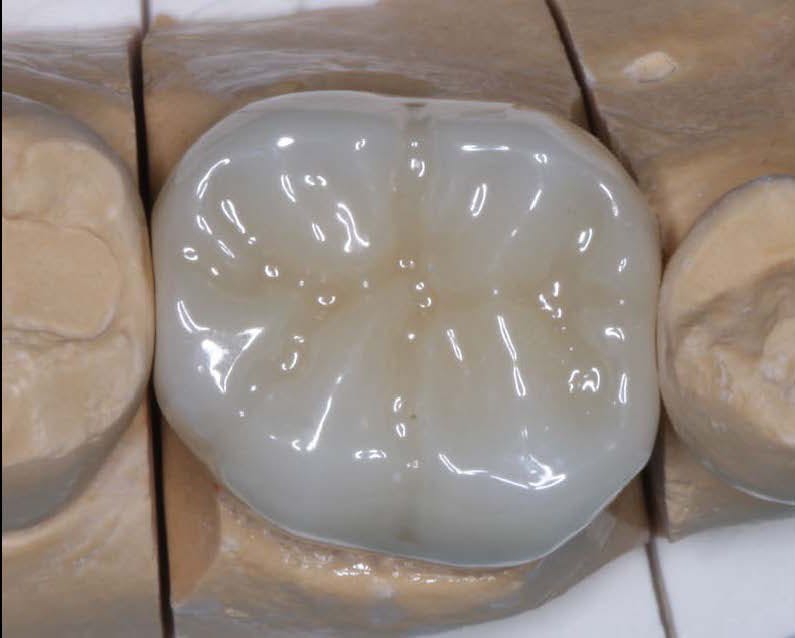
When it comes to restorative dentistry, zirconia crowns have emerged as a superior solution for patients seeking durable and esthetically pleasing dental restorations. Zirconia, a biocompatible ceramic material, offers a range of advantages that make it a popular choice among dentists and patients alike. In this blog post, we will delve into the benefits of zirconia crowns, exploring their strength, aesthetics, and long-term reliability. As a trusted dental lab specializing in zirconia crown fabrication, KC Dental Lab is committed to providing dentists with high-quality restorations that deliver exceptional outcomes.

I. Unparalleled Strength and Durability of Zirconia Crowns:

Zirconia crowns are renowned for their exceptional strength and durability, making them an ideal choice for both posterior and anterior restorations. The inherent properties of zirconia allow for excellent load-bearing capacity, resisting fractures and chipping. This durability ensures that zirconia crowns can withstand the forces of occlusion, providing long-lasting solutions for patients. By partnering with KC Dental Lab, dentists can offer their patients restorations that not only look natural but also offer unmatched strength and reliability.

II. Natural-Looking Aesthetics: Creating Beautiful Smiles:

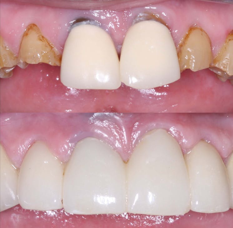
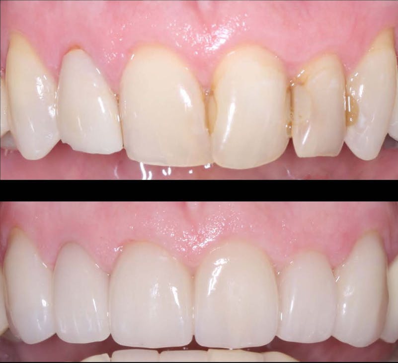
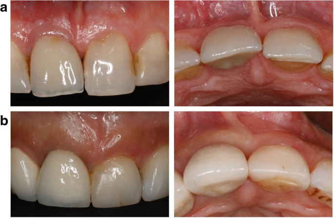
One of the key advantages of zirconia crowns is their ability to replicate the natural appearance of teeth. With their translucent properties and shade-matching capabilities, zirconia crowns blend seamlessly with the surrounding teeth, resulting in esthetically pleasing restorations. This makes zirconia crowns an excellent choice for patients who desire a beautiful smile. KC Dental Lab understands the importance of achieving natural-looking aesthetics and employs advanced techniques to create zirconia crowns that exhibit lifelike characteristics, enhancing patient satisfaction.

III. Biocompatibility and Oral Health Benefits:

Zirconia is a biocompatible material, meaning it is well-tolerated by the human body. Unlike certain metal-based restorations, zirconia crowns do not cause adverse reactions or allergic responses in patients. Additionally, zirconia crowns do not conduct heat or cold, minimizing sensitivity and discomfort for individuals with temperature sensitivity. Furthermore, zirconia crowns have a smooth surface that resists plaque accumulation, promoting better oral hygiene and reducing the risk of periodontal issues. By offering zirconia crowns fabricated by KC Dental Lab, dentists can ensure the oral health and well-being of their patients.

IV. Preserving Tooth Structure: Minimally Invasive Dentistry:

Another significant advantage of zirconia crowns is their ability to preserve natural tooth structure. Due to the high strength of zirconia, minimal reduction of the tooth is required for crown preparation. This conservative approach helps to maintain the integrity of the remaining tooth structure, reducing the need for extensive drilling and preserving healthy tooth enamel. With zirconia crowns, dentists can provide their patients with minimally invasive dentistry, promoting long-term oral health.

V. Long-Term Reliability and Patient Satisfaction:

Zirconia crowns have a proven track record of long-term success, offering patients reliable restorations that stand the test of time. With proper oral hygiene and regular dental visits, zirconia crowns can last for many years, providing patients with a durable and functional solution. The combination of strength, aesthetics, biocompatibility, and preservation of tooth structure contributes to high patient satisfaction. By partnering with KC Dental Lab for zirconia crown fabrication, dentists can ensure that their patients receive restorations that not only meet their functional needs but also exceed their expectations in terms of aesthetics and longevity.
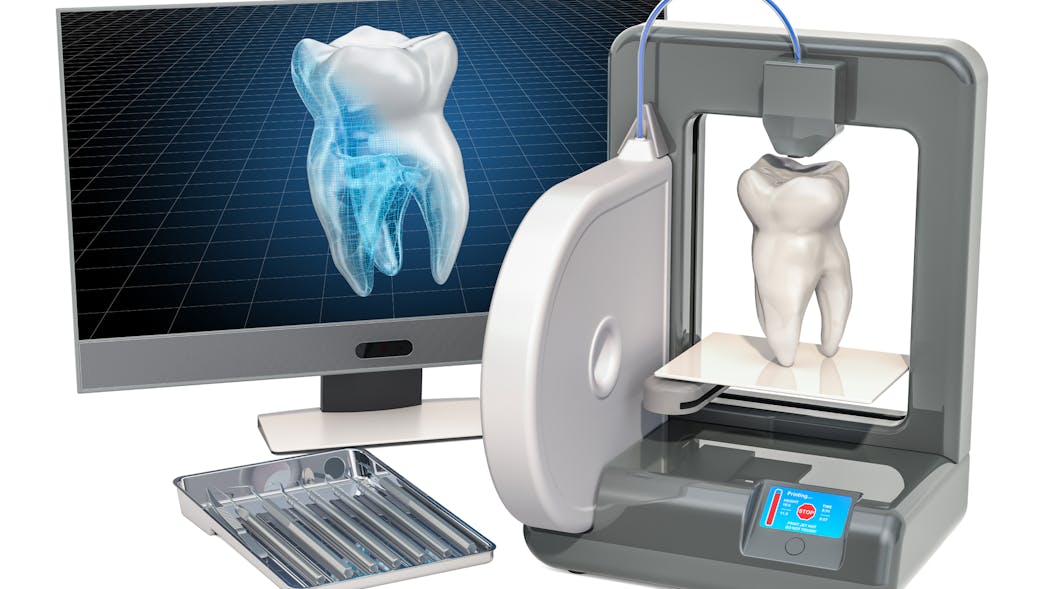
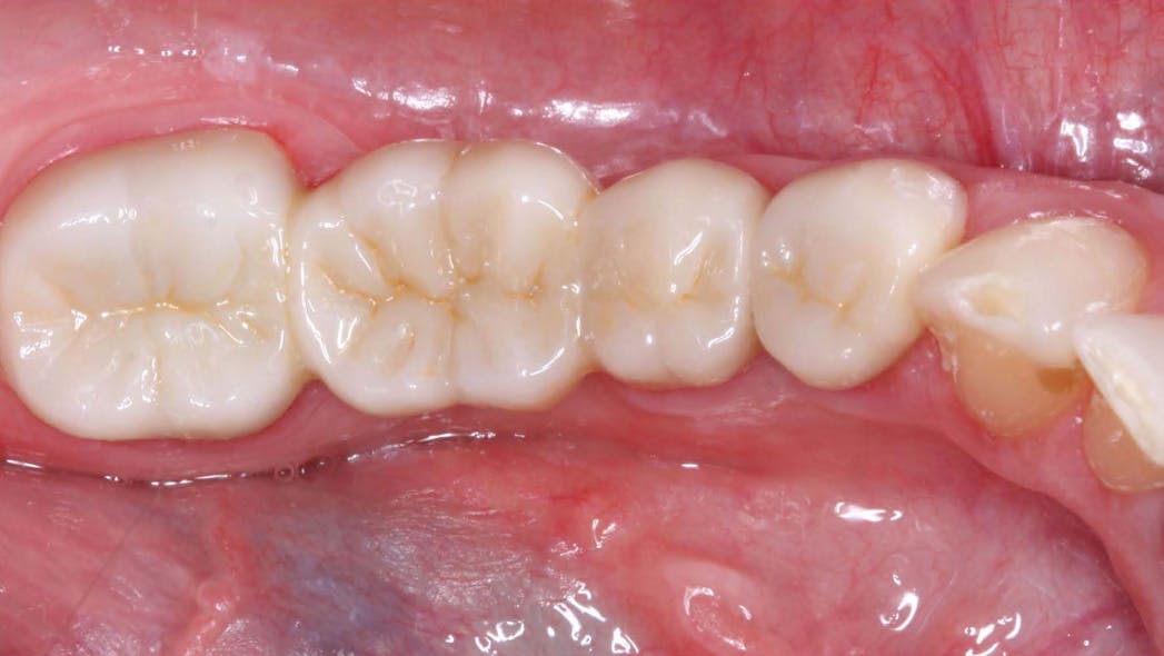
Adhesive Bonding Techniques for Dental Crowns: Advancements in Strength, Durability, and Ease of Use

In the realm of dental crown placement, adhesive bonding techniques have undergone significant advancements, revolutionizing the way dental crowns are attached to natural teeth. These innovative bonding systems and techniques have propelled the field of dentistry forward, offering improved bond strength, enhanced durability, and simplified application methods. In this blog post, we will delve into the latest adhesive bonding techniques for dental crowns, showcasing the advancements in bond strength, durability, and ease of use, ultimately leading to successful and long-lasting restorations.

I. Understanding Adhesive Bonding in Dental Crown Placement:

A. Overview of adhesive bonding and its benefits

B. Importance of a strong bond between the crown and tooth structure

C. Evolution from traditional cementation to adhesive bonding

II. Latest Adhesive Systems for Dental Crown Bonding: A. Resin-based adhesive systems

  1. Introduction to self-etch and total-etch adhesive systems
  2. Advantages and considerations of different adhesive systems B. Bonding agents and primers
  3. Role of bonding agents in enhancing bond strength
  4. Advances in primers for reliable adhesion to different tooth substrates C. Dual-cure and light-cure materials
  5. Benefits of dual-cure materials in challenging clinical situations
  6. Advancements in light-cure materials for simplified application

III. Advancements in Adhesive Bonding Techniques:

A. Selective enamel etching and conditioning

  1. Techniques to preserve enamel and increase bond strength
  2. Advances in selective etching protocols B. Use of adhesive protocols with different substrate conditions
  3. Bonding techniques for vital and non-vital teeth
  4. Strategies for bonding to previously restored teeth or dental implants C. Proper isolation and moisture control
  5. Importance of a dry and isolated field for optimal bond strength
  6. Innovative isolation techniques and materials for effective moisture control

IV. Benefits and Considerations of Adhesive Bonding for Dental Crowns:

A. Improved bond strength and durability

  1. Enhanced retention and resistance to mechanical forces
  2. Reduced risk of microleakage and secondary caries B. Conservative tooth preparation and preservation
  3. Minimal tooth reduction for adhesive bonding compared to traditional methods
  4. Preservation of healthy tooth structure and long-term tooth health C. Simplified application and enhanced user experience
  5. User-friendly techniques for efficient bonding procedures
  6. Time-saving benefits and improved patient comfort

Conclusion: The advancements in adhesive bonding techniques for dental crowns have revolutionized the field of dentistry, offering superior bond strength, enhanced durability, and simplified application methods. With the latest resin-based adhesive systems, bonding agents, and improved techniques, dental professionals can achieve long-lasting and esthetically pleasing restorations while preserving healthy tooth structure. By embracing these advancements, dentists can deliver optimal outcomes, ensuring the success and satisfaction of both clinicians and patients in dental crown placements.

Tips for Choosing a Dental Lab

  1. Look for a dental lab with a good reputation: Do your research and look for a lab that has a good reputation in the industry. Ask for references and check online reviews to get a sense of the lab’s quality and customer service.
  2. Consider the lab’s certifications and accreditation: Look for a lab that is certified by organizations like the National Association of Dental Laboratories (NADL) or the American Dental Association (ADA). These certifications indicate that the lab meets certain standards of quality and professionalism.
  3. Consider the lab’s experience and expertise: Look for a lab that has experience and expertise in the specific dental services you require. For example, if you need implant restorations, look for a lab that specializes in that field.
  4. Check the lab’s turnaround time: Make sure the lab can meet your deadline and turnaround time. A lab that is able to complete your case in a timely manner is important to minimize the time your patient is without a restoration.
  5. Look for a lab that uses modern technology and materials: Look for a lab that uses the latest technology and materials. This will ensure that your patients receive high-quality restorations that are both durable and esthetic.
  6. Look for a lab that offers detailed communication and case tracking: A lab that offers detailed communication and case tracking will help you keep track of your patient’s case and make sure that it is completed on time.
  7. Look for a lab that offers a warranty: Look for a lab that offers a warranty on their work. This will give you peace of mind knowing that if something goes wrong, the lab will take care of it at no additional cost.
  8. Look for a lab that is located near you: Look for a lab that is located near you. This will make it easier to communicate with the lab and drop off and pick up cases.
  9. Look for a lab that offers a variety of services: Look for a lab that offers a variety of services, that way you can always rely on them for any case that you may have
  10. Compare pricing and services: Compare pricing and services of different dental labs and choose the one that offers the best value for your money.

By considering these tips, you will be able to pick a dental lab that meets your needs and delivers high-quality restorations that are both durable and esthetic.

Dental Lab Technicians in the United States

Dental lab technicians play an often underrated role in the US dental care system. It is their job to craft the crowns, bridges, dentures, and other prostheses needed to help an individual recover and restore their smile. It is a highly specialized arrangement that involves craftsmanship, precision, and attention to detail. The US labor force has over 30,000 persons in the dental lab technician field, located from coast to coast and in every state.

The job of tooth technician is not for everyone. Many of those in the field are highly-trained professionals with 10 to 15 years or more experience. Qualifications may include formal education and certifications in dental laboratory technology, dental materials sciences, dental anatomy, and more. It is their commitment to quality and their craftsmanship that ensures a superior product.

For the most part, dental lab technicians are grouped into the category of ‘dental assistant’, but they do perform specialized duties. They first receive a model or request from a dentist, from which they will craft the actual prostheses. During the process, technicians collaborate with the dentist on the design of the prosthesis and also review available materials, advise the dentist on the durability, and make sure it meets the desired aesthetic objectives. Depending on the prosthesis that is being fabricated, the process is often meticulous, requiring many steps and attention to detail.

In addition to prosthetics, technicians may also provide repairs and polishing services for existing prosthetic items. They may also craft retainers, mouth guards, splints, dentures, and similar items. As technology evolves, some technicians are also called upon to perform CAD/CAM operations to embrace the latest fabrication techniques.

Generally, a dental lab technician earns an averagerange of 28,000 to 35,000 US Dollars a year. Some technicians with higher levels of education may earn more, while those with limited experience will likely earn less. It is reported that those technicians working in states like California, Washington, and New York tend to make more money, while the state of Georgia pays their dental lab technicians better than most other states.

Music to their ears? Probably not, but the job of a dental lab technician is essential to the health of everyone in the United States. Without them, there would be no way to produce the prostheses we need to restore smiles and give people their confidence back. They surely deserve our admiration and respect.

Will 3D printing become the new manufacturing standard in dentistry?

Getty Images 1130976572

For almost 40 years,dentistry has been associated with some form of CAD/CAM technology. We have been scanning teeth and models for decades and using CNC technology to mill and manufacture restorations. This process has been purely reductive, meaning a block or ingot is ground or milled down to produce the final restoration. However, with the advent of additive manufacturing—think 3D printing—we are entering a new era of dental manufacturing, and it’s going to be a very exciting time.

Traditional milling of dental restorations began with diamond grinding of glass ceramics. Feldspathic blocks were ground using dual-motor CNC machines to create inlays/onlays and crowns. This process would sometimes take upward of 30–45 minutes. At the time, this was an incredible innovation, but today that amount of time would be simply unacceptable.

Fast-forward to today and the milling processes are light years ahead of where we started. Today, I can send a restoration from my CEREC Primescan, which was designed using only a few clicks, to my Primemill and have a full-contour zirconia restoration manufactured in less than five minutes. It’s incredible to think how much further we can go with the speed, accuracy, and efficiency we have in our current technology. And that is only in the chairside world.

Our laboratory partners have even more incredible technology that allows them not only to manufacture high-quality restorations efficiently, but on a much larger scale. Using large five-axis milling machines, laboratories can manufacture dozens of restorations at once out of a single puck of material. This has helped reduce the overhead for labs and increase their output. The modern dental lab technician may no longer have a CDT degree, but instead a computer science and graphic design background. Design and milling restorations will forever be a computer-guided process.

Within the last five to seven years, the world of 3D printing has exploded onto the dental scene. Formlabs was one of the first manufacturers to target the dental market with its Form2 printer. Using a vat filled with uncured resin, a very detailed laser would systematically cure the resin onto a build platform to create dental models from digital impressions. My first 3D printer would typically take 10–12 hours to manufacture a model, and that was completely acceptable at the time. In fact, it was exciting to finally eliminate alginate, stone, and a model trimmer!

Today, companies such as SprintRay and Dentsply Sirona are creating powerful and innovative 3D printing solutions that are speeding up the manufacturing process by leaps and bounds. On my Primeprint, I can produce a set of models in fewer than 30 minutes, which was unheard of just a few years ago.

With the rapid pace of innovation, research, and development—specifically regarding biocompatible materials and resins—it’s only a matter of time before additive manufacturing becomes the standard in dentistry. It took us almost 40 years to take a milled crown down from 40 minutes to five, and only two to three years to take a 12-hour printing process down to 30 minutes. We are already printing surgical guides and splints and provisionals today. I imagine within a few years, 3D printing of definitive restorations will have a significant place in the market. Regardless, I’m excited to just be along for the ride.  

Editor’s note: This article appeared in the May 2022 print edition of Dental Economics magazine. Dentists in North America are eligible for a complimentary print subscription.

Which crown types are best for what situations?

Figure2

View Image Gallery 

Q: There is enormous confusion in the dental marketplace regarding which indirect restorations should be used and where in the mouth they should be used. The ads are not only confusing, but, in my opinion, they are also too optimistic. The significant commercial information claiming unprecedented success for specific restorations seems unrealistic. What is the state of the art for indirect restorations? What should be used and where in the mouth? How does heavy occlusion factor into the decision? Can optimum esthetics be achieved for all new materials? I would appreciate knowing what Clinical Research (CR) Foundation has found in long-term clinical studies.

A: I answered some of the same questions in “Which crown goes where?” but since then, many changes have occurred, more research is available, and questions keep coming up frequently. Importantly, the research on the various indirect restorations is starting to mature and provide some answers. In this article, I will give you a status report on the most-used crown types and their current success in CR/TRAC (Technologies in Restoratives and Caries Research Division of the CR Foundation) in vivo research. The information is divided into several locations in the mouth along with my suggestions as to the different strength and esthetic needs for those locations.


Also by Dr. Christensen:

Increasing practice activity

Are endodontic posts really necessary?


The following overall statements relate to my answers.

  • It is reported by large US dental labs that over 74% of indirect restorations are for single teeth.1
  • Large labs report that ceramic indirect restorations currently comprise over 90% of the total indirect restorations made. The majority of those indirect restorations are milled from one of the zirconia variations, some are milled or pressed lithium disilicate, and a small number are conventional porcelain-fused-to-metal (PFM), or polymer.1
  • A very conservative estimate is that about one-third of the adult population have either grinding or clenching bruxism, a highly important characteristic for restoration selection.
Figure 1: Class 5, tetragonal, 3Y zirconia—the original BruxZir has revolutionized the dental profession. It is now well proven to be the strongest and most durable ceramic restoration in dentistry, but color challenges still exist.

It can be concluded from the previous statements that different types of indirect restorations are present and that their various physical and esthetic characteristics relate to where they should be used. It is also important to note that additional long-term research is needed to confirm some of my suggestions.

Molars

Optimum strength is available by using class 5, 3Y, tetragonal zirconia (figure 1). This is the original BruxZir formulation, now available from many Glidewell laboratories. Similar products are produced by other companies under other names. It is often called LT (“low translucency”) zirconia by dentists and labs. The strength and durability of this zirconia category have been confirmed through 11 years of in vivo research by the TRAC Division.

Figure 2: A three-unit fixed prosthesis and a separate single crown milled from class 5 zirconia with a thin layer of veneering ceramic/glaze to make it an acceptable color. However, the thin veneer/glaze will eventually wear off the occlusal surface.

But, as most dentists know, this zirconia category has less-than-desirable esthetic qualities unless coated with layering ceramic or stained in the presintered zirconia stage (figures 2 and 3). Some dentists do not object to the unmodified color of this zirconia category for molars since it is not usually visible in the posterior of the mouth.

This zirconia formulation is well proven and has had unprecedented clinical success and lack of breakage. However, labs and manufacturers primarily promote the more esthetic forms of zirconia, identified as class 4 cubic zirconia. It is often described as HT (high translucency), or esthetic zirconia. This form of zirconia has lower strength than class 5 zirconia, and it still lacks long-term research for use in high-strength needs.

Figure 3: A four-unit fixed prosthesis milled from class 5 zirconia placed in a patient with extreme bruxing with pigment placed on the zirconia in the presintered stage to make the color acceptable.

Currently, clinical research has mixed results, indicating promise for class 4 zirconia formulations but also some potential challenges. Since this formulation has been available for only a few years, you and your peers are doing much of the observational clinical research on these materials in your practices. Long-term clinical research is still needed to validate the use of this zirconia form for molars, bruxing patients, long-span fixed prostheses, and other high-strength clinical needs.

Premolars

IPS e.max (lithium disilicate) is classified as a class 3 ceramic restoration. It is very well proven for single premolar restorations by both controlled studies and millions of such restorations placed internationally. As you know, it has unprecedented high esthetic qualities and strength (figures 4 and 5).

Figure 4: IPS e.max can cover even dark-colored teeth if it is at least 1.0 mm thick in all axial aspects. The left lateral incisor replacement is an implant. It has a thin metal opaqued coping placed over the implant, making the color of the class 3 ceramic restoration (lithium disilicate) acceptable.

For nonbruxers, IPS e.max can be used safely for single premolars and select three-unit fixed prostheses involving both premolars and anterior teeth. It is suggested that at least 1.0 mm of IPS e.max thickness is present on all axial walls and 1.5–2.0 mm thickness on the occlusal surface for optimal strength. However, more fractures have been reported on multiple-unit lithium disilicate fixed prostheses in the premolar to anterior area than on single teeth.

Should IPS e.max be used on premolars in bruxing patients? Some practitioners are using it in bruxing situations because of its great success in nonbruxers. The only alternatives are porcelain-fused-to-metal (PFM) or class 4 zirconia. Selecting an appropriate restoration for a bruxing patient requires that the dentist have personal knowledge of the patient, the anticipated occlusal loading, and 

Figure 5: The strength and esthetics of IPS e.max for single crowns is unexcelled by other materials.

any esthetic needs.

Should class 4 cubic zirconia restorations be used for premolars? These modified zirconia forms are being highly promoted for such situations. Clinical observation by CR evaluators has been promising, but those observations are only short term. Developing challenges have already been noted in some brands by CR’s in vitro microscopic research, indicating the use of class 4 zirconia with caution until additional long-term research is available.

At present, IPS e.max is a well-proven product for single premolars and select three-unit fixed prostheses involving premolars.

In situations involving high-strength needs, such as in bruxing patients, color-modified class 5, 3Y zirconia is still a more-proven concept. Additional clinical research will determine if class 4 zirconia will be adequate for patients who are bruxers.

Anterior teeth

Anterior teeth usually have the least need for strength. The statements on premolars apply directly to anterior teeth, but esthetic acceptability is more important in the anterior area of the mouth.

If the restoration is for single teeth and the patient is not a bruxer, IPS e.max currently is the optimum restoration.

If a three-unit (or larger) fixed prosthesis is needed, color-modified class 5, tetragonal, 3Y zirconia may be optimum, especially for a bruxing patient, but such zirconia requires an artist/technician to achieve the best esthetics.

Should class 4 cubic zirconia be considered for anterior three-unit (or larger) fixed prostheses? The same challenges are present as those for premolars. More long-term research is needed. How long? At least several years of research on many brands. That research is beginning to come forth, but there are still numerous questions. If using class 4 zirconia for anterior restorations, I suggest observing the restorations carefully with high-power loupes at each recall appointment. Look for pits, minor cracks, excessive wear on opposing teeth, or other maladies. These challenges have been seen on some class 4 zirconia brands in preliminary research. It is our hope that class 4 zirconia brands will soon prove themselves in clinical research. In the meantime, be observant and cautious.

Summary

Significant confusion is present about what type of indirect restoration is best for specific situations. Current evidence, both scientific and observational, support the use of class 5, tetragonal, 3Y zirconia. However, this formulation has esthetic challenges that must be overcome. Class 4 cubic-containing zirconia has many formulations. Many brands are currently proving themselves, but more years will be necessary for that proof to be solidified.

IPS e.max is well proven for near universal use in nonbruxers and limited use in bruxers. In the meantime, don’t forget the more than 120 years of success dentistry has had with cast-gold alloy and the more than 65 years of success with porcelain-fused-to-metal.

The immediate future appears to point to continued and expanded use of zirconia indirect restorations with a slow reduction in the use of the excellent, well-proven IPS e.max.  

Reference

  1. Based on data from Glidewell Labs.

Author’s note: The following educational materials from Practical Clinical Courses offer further resources on this topic for you and your staff.

One-hour videos:

  • Cementing Restorations—Proven and Successful (Item no. 1921)
  • Impressions Can Be Simple and Predictable (Item no. 1922)

Two-day hands-on courses in Utah:

  • Restorative Dentistry 1—Restorative/Esthetic/Preventive with Dr. Gordon Christensen
  • Faster, Easier, Higher Quality Dentistry with Dr. Gordon Christensen

For more information, visit pccdental.com or contact Practical Clinical Courses at (800) 223-6569.

Editor’s note: This article originally appeared in the February 2022 print edition of Dental Economics.

The influence of soft-tissue volume grafting on the maintenance of peri-implant tissue health and stability

Abstract

Background

To investigate the influence of soft-tissue volume grafting employing autogenous connective tissue graft (CTG) simultaneous to implant placement on peri-implant tissue health and stability.

Material and methods

This cross-sectional observational study enrolled 19 patients (n = 29 implants) having dental implants placed with simultaneous soft-tissue volume grafting using CTG (test), and 36 selected controls (n = 55 implants) matched for age and years in function, who underwent conventional implant therapy (i.e., without soft-tissue volume grafting). Clinical outcomes (i.e., plaque index (PI), bleeding on probing (BOP), probing depth (PD), and mucosal recession (MR)) and frequency of peri-implant diseases were evaluated in both groups after a mean follow-up period of 6.15 ± 4.63 years.

Results

Significant differences between test and control groups at the patient level were noted for median BOP (0.0 vs. 25.0%; p = 0.023) and PD scores (2.33 vs. 2.83 mm; p = 0.001), respectively. The prevalence of peri-implant mucositis and peri-implantitis amounted to 42.1% and 5.3% in the test and to 52.8% and 13.9% in the control group, respectively.

Conclusion

Simultaneous soft-tissue grafting using CTG had a beneficial effect on the maintenance of peri-implant health.

Introduction

A major goal of implant therapy is to ensure long-term peri-implant tissue health and create appealing esthetics. To obtain these therapeutic endpoints, soft-tissue grafting procedures performed either simultaneously with or after implant placement have become an indispensable part of contemporary implant dentistry [1].

From a biological point of view, a lack of or reduced height (< 2 mm) of keratinized mucosa (KM) around the implants was shown to jeopardize self-performed oral hygiene measures, which subsequently increased the likelihood of soft-tissue inflammation [12]. As a consequence, soft-tissue grafting procedures aimed at increasing keratinized tissue have been shown to markedly improve peri-implant soft-tissue inflammatory conditions and were associated with higher marginal bone levels compared to the control sites [3]. Moreover, from an esthetic perspective, the presence of KM > 2 mm was demonstrated to be a preventive measure for the occurrence of peri-implant soft-tissue dehiscences [4].

Changes in peri-implant soft-tissue height, particularly on the facial aspect, are a critical factor that may compromise the overall esthetic result of implant-supported restoration [5]. A thin mucosa (also known as a soft-tissue biotype) at the time of implant installation was found to be a crucial component that correlated with facial soft-tissue recession [6,7,8]. In fact, to attenuate the undesirable changes of the soft-tissue margin, soft-tissue volume augmentation at the time of implant placement was also suggested as a preventive measure [910]. On the contrary, currently available data evaluating procedures to increase mucosal thickness did not show any significant effects on bleeding scores, but higher interproximal marginal bone levels over time when compared with control sites [1]. Due to a lack of reporting, an evaluation of the prevalence of peri-implant disease was not feasible [1].

Therefore, the aim of the present cross-sectional analysis was to assess the influence of soft tissue volume grafting on the peri-implant tissue health and stability.

Materials and methods

The present investigation was designed as an observational, cross-sectional case–control study evaluating the clinical treatment outcomes of implants inserted simultaneously with (test group) and without (control group) soft-tissue volume augmentation. All patients had received the same implant brand (Ankylos®, Dentsply Sirona Implants, Hanau, Germany) in a single university clinic (Department of Oral Surgery and Implantology, Goethe University, Frankfurt) and were recruited during their yearly maintenance visits.

Patients were included in the study once they were informed about the investigation procedures and gave their written informed consent. The procedures in the present study were in accordance with the Declaration of Helsinki, as revised in 2013, and the study protocol was approved by the local ethics committee (registration number: 78/18).

Patient selection criteria

The following inclusion criteria were applied for patient selection:

– Patients with > 18 years of age rehabilitated with at least one Ankylos® implant;

– Patients with treated chronic periodontitis and proper periodontal maintenance care;

– Non-smokers, smokers and former smokers;

– A good level of oral hygiene as evidenced by a plaque index (PI) < 1 at the implant level; and

– Attendance of yearly routine implant maintenance appointment.

Patients were excluded for the following conditions: the presence of combined endodontic–periodontal lesions; systemic diseases that could influence the outcome of the therapy, such as diabetes (HbA1c > 7), osteoporosis and antiresorptive therapy; a history of malignancy, radiotherapy, chemotherapy, or immunodeficiency; and pregnancy or lactation at the last follow-up.

Surgical protocol

Soft-tissue biotype was assessed preoperatively based on the probe’s transparency at the mid-facial aspect and categorized as thin when the probe was visible and thin when it was not visible. Two-piece platform-switched implants were placed 2–3 mm subcrestally according to the manufacturer’s surgical protocol. Implants in the control group exhibited a thick soft-tissue biotype and therefore underwent a conventional placement protocol (i.e., without soft-tissue volume grafting; Fig. 1a).

figure1
Fig. 1

Implants in the test group presented with a thin soft-tissue biotype, and therefore, a connective tissue graft (CTG) harvested from the hard palate was simultaneously applied on the facial aspect via tunneling technique (Fig. 1b and Fig. 2). All surgeries were performed by one experienced oral surgeon (PP).

figure2
Fig. 2

Implant and implant-site characteristics

The following study variables were assessed for the test and control implant sites: (1) implant age (i.e., defined as time after implant placement), (2) implant location in the upper jaw, and (3) implant diameter.

Clinical measurements

The following clinical parameters were registered at each implant site using a periodontal probe: (1) plaque index (PI) (Löe et al., 1967); (2) bleeding on probing (BOP)—measured as presence/absence; (3) probing depth (PD)—measured from the mucosal margin to the probable pocket; (4) mucosal recession (MR)—measured from the restoration margin to the mucosal margin; and (5) keratinized mucosa (KM) (mm)—measured on the buccal aspects of the implants.

PI, BOP, PD, and MR measurements were performed at six aspects per implant site: mesiobuccal (mb), midbuccal (b), distobuccal (db), mesiooral (mo), midoral (o), and distooral (do). KM measurement was performed at three aspects per implant site: mesiobuccal (mb), midbuccal (b), and distobuccal (db).

The presence of peri-implant diseases at each implant site was assessed as follows [11]:

  • Peri-implant mucositis defined as the presence of BOP and/or suppuration with on gentle probing with or without increased PDs compared to previous examinations and an absence of bone loss beyond crestal bone level changes resulting from initial bone remodeling.
  • Peri-implantitis defined as the presence of BOP and/or suppuration on gentle probing, increased PDs compared to previous examination, and the presence of bone loss beyond crestal bone level changes resulting from initial bone remodeling.

Radiographs (i.e., panoramic) were just taken when clinical signs suggested the presence of peri-implant tissue inflammation (i.e., the presence of BOP). To estimate the bone level changes at the respective implant sites, these radiographs were compared with those taken following the placement of the final prosthetic reconstruction (i.e., baseline).

Investigators meeting and calibration

Prior to the start of the study, a calibration meeting was held with each examiner (KO, AB, AR) to standardize (pseudonymous) data acquisition and the assessment of study variables. For the calibration of the examiners, double measurements were performed with a 5-min interval of the assessed clinical parameters in 5 patients with a total of 15 implants. The calibration was acceptable when repeated measurements were similar > 95% level. The documentation of demographic study variables, implant sites’ characteristics, and clinical measurements were documented using a generated standardized data extraction template.

Statistical analysis

The statistical analysis was performed using a commercially available software program (SPSS Statistics 27.0: IBM Corp., Ehningen, Germany). Descriptive statistics (means, standard deviations, medians and 95% confidence intervals) were calculated for mPI, BOP, PD, and MR values. The analysis was performed at the patient and implant levels. The data were tested for normality by means of the Shapiro-Wilk test. Comparisons of clinical parameters between the test and control groups were performed by employing the Mann-Whitney U test. Linear regression analyses were used to depict the relationship between mean BOP, PD, and MR values and KM scores. The alpha error was set at 0.05.

Results

Patient and implant sites’ characteristics

The test group included 19 patients (13 women and 6 men) with a total of 29 implants, whereas the control group included 36 patients (20 women and 16 men) with a total of 55 implants. Mean patient age in the test and control groups was 46.24 ± 18.48 and 62.21 ± 14.41 years, respectively. The mean implant functioning time was 4.16 ± 2.06 years for the test group and 7.19 ± 5.25 years for the control group. All implants in the test group revealed a diameter of 3.5 mm with an equal distribution between all regions investigated. In the control group, the most frequent diameter was also 3.5 mm (85.5%), with a predominant implant location in the region of the lateral and central incisors (Table 1).Table 1 Patient and implant site characteristicsFull size table

Clinical measurements

The results of the clinical measurements are presented in Table 2. In general, test and control groups were commonly characterized by low median PI scores at both patient (0.00 vs. 0.21; p = 0.093) and implant levels (0.17 vs. 0.17), respectively.Table 2 Clinical parameters (mean ± SD, median and 95% CI)Full size table

Marked differences between test and control groups were noted for median BOP scores, reaching statistical significance at the patient level (0.0 vs. 25.0%; p = 0.023).

Similarly, the test group was associated with markedly lower median PD values at both patient (2.33 vs. 2.83 mm; p = 0.001) and implant levels (2.33 vs. 2.83 mm), respectively.

Both groups revealed comparable median MR values at both patient (0.0 vs. 0.0 mm; p = 0.76) and implant levels (0.0 vs. 0.0 mm), respectively (Table 2).

Prevalence of peri-implant diseases

The frequency distribution of peri-implant diseases in the test and control groups at patient and implant levels is summarized in Tables 3 and 4.Table 3 Prevalence of peri-implant disease (patient level)Full size tableTable 4 Prevalence of peri-implant disease (implant level)Full size table

According to the given case definitions, 66.7% of the patients in the control group and 47.4% of the patients in the test group were diagnosed with peri-implant diseases. In the test group, the prevalence of peri-implant mucositis and peri-implantitis amounted to 42.1% and 5.3%. In the control group, the corresponding values were 52.8% and 13.9%, respectively (Table 3).

At the implant level, the prevalence of peri-implant mucositis and peri-implantitis amounted to 44.8% and 3.4% in the test group, and 52.7 and 9.1% in the control group, respectively (Table 4).

Regression analysis

Cross-tables depicting selected independent variables (PD, MR, and BOP values) and local factors (i.e., KM and Implant age) in both test and control groups are summarized in Tables 5and 6.Table 5 Test group (n = 29 implants). Cross-tables of BOP/PD/MR values and (1) KM and (2) implant age (months)Full size tableTable 6 Control group (n = 55 implants). Cross-tables of BOP/PD/MR values and (1) KM and (2) implant age (months)Full size table

In the test group, the linear regression analysis failed to reveal any significant correlations between KM and the independent variables investigated.

In the control group, a significant correlation was noted between KM and MR values (R2 = 0.155; B = − 0.072; p = 0.003) (Tables 5 and 6).

Discussion

The present cross-sectional analysis aimed at investigating the influence of soft-tissue volume grafting employing autogenous CTG simultaneous to implant placement on peri-implant tissue health and stability. Based on the clinical parameters investigated, it was noted that the patients in the test group revealed significantly lower BOP and PD scores when compared with those of the control group. This was associated with a lower prevalence of peri-implant diseases, particularly of patients diagnosed for peri-implantitis. In this context, it must be emphasized that the latter assessment was based on recently established case definitions and considered previous examination data [11].

Basically, the present results do not confirm the findings of a recent systematic review and meta-analysis, since soft tissue grafting procedures by means of CTG were not associated with any significant differences in BOP or PD values as compared to control treatments [1]. The analysis was based on a total of 6 randomized (n = 2)/controlled clinical (n = 4) studies reporting on a total of 260 systemically and periodontally healthy patients over a mean follow-up period of 57 months [912,13,14,15,16]. Except for one study [17], the implants were placed immediately and soft tissue grafting was accomplished either at implant placement [91215], or after a healing period of 3 months [131416]. At test sites, the range of mean BOP values was 20–35% at baseline and amounted to 20–56% at follow-up [14,15,16,17]. The corresponding values at control sites were 21–40% at baseline and 33–46% at follow-up [1]. A total of five studies [913,14,15,16] failed to identify any significant effects of soft-tissue volume grafting on mean PD values. In particular, at test sites, the range of mean PD values was 2.50–3.45 mm at baseline and amounted to 3.67–4.09 mm at follow-up. At control sites, these values were 2.50–3.20 mm at baseline and 3.20–3.97 mm at follow-up [1]. One study focusing on immediate implant placement with simultaneous soft-tissue volume grafting reported on significantly lower PD values at test sites when compared with control sites [12].

The meta-analysis failed to reveal any significant differences in either plaque, BOP, or PD scores (i.e., changes or endpoint values) between test and control groups. However, significantly less marginal bone loss over time was observed with the use of CTG [n = 2; WMD = 0.110; 95% CI (0.067; 0.154); p < 0.001] when compared to sites without grafting [1].

The discrepancy noted between the present analysis and the aforementioned systematic review may, at least in part, be explained by the fact that the included studies [912,13,14,15,16] did not consider BOP or PD as primary outcomes measures. Accordingly, the power of these studies may not have been sufficient to rule out potential differences between groups. Moreover, it needs to be emphasized that none of the evaluated studies [912,13,14,15,16] used case definitions for the evaluation of the occurrence of peri-implant diseases [1].

The present study did not consider to routinely take radiographs during follow-up, but just limited the indication to those patients exhibiting clinical signs of peri-implant tissue inflammation [18]. Accordingly, the influence of soft-tissue volume grafting procedures on marginal bone level changes could not be assessed.

When further evaluating the present data, it was also noted that, in contrast to implants of the test group, control sites revealed a significant correlation between KM and MR values. In this context, it must be emphasized that a major drawback of the present study was the lack of a quantification of the horizontal mucosal thickness (i.e., biotype) during follow-up. That was due to the fact that the assessment of the biotype is challenging at diseased implant sites, since the inflammatory lesion is inevitably associated with an increase in mucosal thickness [19]. As a consequence of the notable prevalence of peri-implant diseases in both groups, it may have been impossible to estimate true changes of the biotype during follow-up.

Nevertheless, the findings of the regression analysis corroborate the results of previous studies also indicating that at implant sites exhibiting a healthy peri-implant mucosa, a thick tissue biotype was associated with a lower frequency of facial soft-tissue recessions (i.e., MR values) over time when compared with sites exhibiting a thin biotype [620].

In conclusion and within its limitations, the present study has indicated that simultaneous soft-tissue grafting using CTG had a beneficial effect on the maintenance of peri-implant health.

Obreja, K., Ramanauskaite, A., Begic, A. et al. The influence of soft-tissue volume grafting on the maintenance of peri-implant tissue health and stability. Int J Implant Dent7, 15 (2021). https://doi.org/10.1186/s40729-021-00295-1

Zircon Lab is America’s leading dental lab. We are partnered with dental offices nationwide and are consistently growing. As America’s highest quality dental lab with the most competitive pricing, the highest caliber of product, expert craftsmanship, and fastest delivery, we set the dental industry standard. After choosing Zircon Lab to be your dental lab of choice, you can trust our dental product will be unmatched by any competitors.

Zircon Lab with Border_White

Choose The Right Dental Lab, The First Time

    Zircon Lab, LLC specializes in high end Zirconia (Bruxer), Aesthetic High Translucent Full Zirconia Crowns for Anteriors, e.max, and implants. All of our restorations are 100% made in the USA and we are the only dental lab to have an unconditional, 100% lifetime warranty.

Contact Info

8060 Reeder St. | Overland Park, KS 66214

+1 888.880.3383
contact@zirconlab.com

Monday - Friday: 8:00 am - 5:00 pm CST
Saturday & Sunday: Closed

Copyright 2023 Zircon Lab, LLC ©  All Rights Reserved