Dentist,In,Coverall,Pointing,At,Green,Screen,Display,During,Coronavirus

Cloud-Based Dental Software Helps Practices Adapt to The New Normal

Since the American Dental Association began tracking the impact of COVID-19 on dental practices in March 2020, there has been a slow and steady climb towards normalcy. 

But we’re not there yet, based on research from January 2021. According to the survey, 32.3% of practices were open and back to business as usual. 66.7% were open but experiencing lower patient volume than usual. Clearly, the road ahead could be long for many practices.

During the early days of the pandemic, many dentists felt overwhelmed as they weighed their options, seeking information on the new regulations for PPE, FFCRA, FMLA, and more. When they received approval to re-open, they had to determine how best to keep their patients, staff, and themselves safe while running a profitable business. This entailed reevaluating their entire workflow, including the practice management software.

Cloud-Based Dental Practice Management Software Front and Center

Unlike other medical segments, dentistry has been slower to adopt cloud-based software. While it is fair to say that the majority of dentists believe that the cloud is the future, approximately 85% were still hanging on to their server-based software as COVID-19 hit in Spring 2020. With limited access to their system and patient data during the shutdown, more practices than ever considered the benefits of the cloud, and many made the move.

Curve Hero™, Curve Dental’s cloud-based dental practice management software, experienced a significant spike in product demos during COVID-19. During the first few months of the pandemic, nearly three times as many practices moved to Curve’s cloud-based platform than normal. In February 2021, Curve announced that over 33,000 dental professionals used Curve Hero, far more than any other cloud-based provider.

Remote Access to Data Helped Curve Hero Customers Get a Jump Start on Recovery

Early on, Curve customers found how much easier it was to open their practice’s doors to their patients while using the Curve Hero platform. Practices made digital forms available to patients whose information automatically went directly into their Curve Hero database. Office staff informed patients of the practice’s COVID-19 protocols in advance of appointments which increased confidence in their commitment to keeping everyone safe. Billing went through the Patient Portal, eliminating the need to handle and return credit cards at the front desk. This “low-touch/no-touch” experience made a very challenging process far more manageable and safe than practices using traditional server-based systems.

What New Customers Said After Switching to Curve Hero  

Going into the demo, dental professionals knew that cloud-based software allowed them much easier access to patient data than their server-based system. But they discovered many more benefits available with Curve Hero. Typically, dentists and office managers are reluctant to change software because of the anticipated disruption to their practice due to the data conversion process, a potential lengthy learning curve, and time-consuming training.

During product evaluations, they learned that Curve makes it significantly easier to switch software by having proven processes in place to make the transition as smooth as possible. Curve collaborates with the dental office from start to finish — during the initial setup through data review and final assembly. Curve has successfully completed more than 4,000 data, file, and image conversions from well over 90 practice management software products, both server-based and cloud-based. Watch Dr. Jesse Ritter explain his Curve Hero conversion experience in this video.

Web-based training means your team doesn’t have to travel. Staff adopts the software quickly because Curve separates each training session into small digestible bites. Plus, your staff has access to Curve Community, a rich library of information to remind them of what they learned or act as a quick training refresher.

Curve’s New Patient Engagement Feature Makes Practices Even More Productive

Recently, Curve added Curve GRO™, a patient engagement feature that simplifies and streamlines communications by having everything in a single system. Powered by a robust campaign engine, Curve GRO automatically manages patient reminder campaigns and updates the schedule when the patient confirms. For patients who may need to change their appointment or ask questions, GRO delivers 2-way conversational texting. For patients who do not respond to the reminder campaigns, GRO can automatically create tasks in the Smart Action List, allowing the staff to collaborate in real-time to triage patient outreach. A rules-based campaign engine combined with the Smart Action List is significant for dentistry because it creates automation, enforces best practices as determined by the practice administrator, and delivers an auditable trail of all activity that occurs.

Disasters Aren’t Planned. They Just Happen.

Well before COVID-19, dental practices have had to deal with unexpected events like fires, floods, data breaches, and more. If your data is contained on a server in your office and disaster strikes, you could be out of luck. There are so many good reasons to move your practice to the cloud starting with protecting your data. In addition, as we learned during the early stages of COVID-19 with the mandatory office shutdown, the ability to access data remotely to triage dental emergencies and manage rescheduling, billing, and payments were extremely beneficial to Curve customers and their patients. The cloud is by far your best option to protect your business from the unexpected.

About Curve:

Founded in 2004, Curve Dental provides web-based dental software and related services to dental practices within the United States and Canada. The company strives to make dental software less about computers and more about user experience. Curve’s creative thinking can be seen in the design of software that is easy to use and built only for the cloud. Visit www.curvedental.com for more information.

Zircon Lab is America’s leading dental lab. We are partnered with dental offices nationwide and are consistently growing. As America’s highest quality dental lab with the most competitive pricing, the highest caliber of product, expert craftsmanship, and fastest delivery, we set the dental industry standard. After choosing Zircon Lab to be your dental lab of choice, you can trust our dental product will be unmatched by any competitors.

The influence of soft-tissue volume grafting on the maintenance of peri-implant tissue health and stability

Abstract

Background

To investigate the influence of soft-tissue volume grafting employing autogenous connective tissue graft (CTG) simultaneous to implant placement on peri-implant tissue health and stability.

Material and methods

This cross-sectional observational study enrolled 19 patients (n = 29 implants) having dental implants placed with simultaneous soft-tissue volume grafting using CTG (test), and 36 selected controls (n = 55 implants) matched for age and years in function, who underwent conventional implant therapy (i.e., without soft-tissue volume grafting). Clinical outcomes (i.e., plaque index (PI), bleeding on probing (BOP), probing depth (PD), and mucosal recession (MR)) and frequency of peri-implant diseases were evaluated in both groups after a mean follow-up period of 6.15 ± 4.63 years.

Results

Significant differences between test and control groups at the patient level were noted for median BOP (0.0 vs. 25.0%; p = 0.023) and PD scores (2.33 vs. 2.83 mm; p = 0.001), respectively. The prevalence of peri-implant mucositis and peri-implantitis amounted to 42.1% and 5.3% in the test and to 52.8% and 13.9% in the control group, respectively.

Conclusion

Simultaneous soft-tissue grafting using CTG had a beneficial effect on the maintenance of peri-implant health.

Introduction

A major goal of implant therapy is to ensure long-term peri-implant tissue health and create appealing esthetics. To obtain these therapeutic endpoints, soft-tissue grafting procedures performed either simultaneously with or after implant placement have become an indispensable part of contemporary implant dentistry [1].

From a biological point of view, a lack of or reduced height (< 2 mm) of keratinized mucosa (KM) around the implants was shown to jeopardize self-performed oral hygiene measures, which subsequently increased the likelihood of soft-tissue inflammation [12]. As a consequence, soft-tissue grafting procedures aimed at increasing keratinized tissue have been shown to markedly improve peri-implant soft-tissue inflammatory conditions and were associated with higher marginal bone levels compared to the control sites [3]. Moreover, from an esthetic perspective, the presence of KM > 2 mm was demonstrated to be a preventive measure for the occurrence of peri-implant soft-tissue dehiscences [4].

Changes in peri-implant soft-tissue height, particularly on the facial aspect, are a critical factor that may compromise the overall esthetic result of implant-supported restoration [5]. A thin mucosa (also known as a soft-tissue biotype) at the time of implant installation was found to be a crucial component that correlated with facial soft-tissue recession [6,7,8]. In fact, to attenuate the undesirable changes of the soft-tissue margin, soft-tissue volume augmentation at the time of implant placement was also suggested as a preventive measure [910]. On the contrary, currently available data evaluating procedures to increase mucosal thickness did not show any significant effects on bleeding scores, but higher interproximal marginal bone levels over time when compared with control sites [1]. Due to a lack of reporting, an evaluation of the prevalence of peri-implant disease was not feasible [1].

Therefore, the aim of the present cross-sectional analysis was to assess the influence of soft tissue volume grafting on the peri-implant tissue health and stability.

Materials and methods

The present investigation was designed as an observational, cross-sectional case–control study evaluating the clinical treatment outcomes of implants inserted simultaneously with (test group) and without (control group) soft-tissue volume augmentation. All patients had received the same implant brand (Ankylos®, Dentsply Sirona Implants, Hanau, Germany) in a single university clinic (Department of Oral Surgery and Implantology, Goethe University, Frankfurt) and were recruited during their yearly maintenance visits.

Patients were included in the study once they were informed about the investigation procedures and gave their written informed consent. The procedures in the present study were in accordance with the Declaration of Helsinki, as revised in 2013, and the study protocol was approved by the local ethics committee (registration number: 78/18).

Patient selection criteria

The following inclusion criteria were applied for patient selection:

– Patients with > 18 years of age rehabilitated with at least one Ankylos® implant;

– Patients with treated chronic periodontitis and proper periodontal maintenance care;

– Non-smokers, smokers and former smokers;

– A good level of oral hygiene as evidenced by a plaque index (PI) < 1 at the implant level; and

– Attendance of yearly routine implant maintenance appointment.

Patients were excluded for the following conditions: the presence of combined endodontic–periodontal lesions; systemic diseases that could influence the outcome of the therapy, such as diabetes (HbA1c > 7), osteoporosis and antiresorptive therapy; a history of malignancy, radiotherapy, chemotherapy, or immunodeficiency; and pregnancy or lactation at the last follow-up.

Surgical protocol

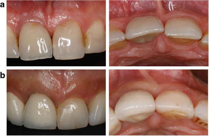
Soft-tissue biotype was assessed preoperatively based on the probe’s transparency at the mid-facial aspect and categorized as thin when the probe was visible and thin when it was not visible. Two-piece platform-switched implants were placed 2–3 mm subcrestally according to the manufacturer’s surgical protocol. Implants in the control group exhibited a thick soft-tissue biotype and therefore underwent a conventional placement protocol (i.e., without soft-tissue volume grafting; Fig. 1a).

figure1
Fig. 1

Implants in the test group presented with a thin soft-tissue biotype, and therefore, a connective tissue graft (CTG) harvested from the hard palate was simultaneously applied on the facial aspect via tunneling technique (Fig. 1b and Fig. 2). All surgeries were performed by one experienced oral surgeon (PP).

figure2
Fig. 2

Implant and implant-site characteristics

The following study variables were assessed for the test and control implant sites: (1) implant age (i.e., defined as time after implant placement), (2) implant location in the upper jaw, and (3) implant diameter.

Clinical measurements

The following clinical parameters were registered at each implant site using a periodontal probe: (1) plaque index (PI) (Löe et al., 1967); (2) bleeding on probing (BOP)—measured as presence/absence; (3) probing depth (PD)—measured from the mucosal margin to the probable pocket; (4) mucosal recession (MR)—measured from the restoration margin to the mucosal margin; and (5) keratinized mucosa (KM) (mm)—measured on the buccal aspects of the implants.

PI, BOP, PD, and MR measurements were performed at six aspects per implant site: mesiobuccal (mb), midbuccal (b), distobuccal (db), mesiooral (mo), midoral (o), and distooral (do). KM measurement was performed at three aspects per implant site: mesiobuccal (mb), midbuccal (b), and distobuccal (db).

The presence of peri-implant diseases at each implant site was assessed as follows [11]:

  • Peri-implant mucositis defined as the presence of BOP and/or suppuration with on gentle probing with or without increased PDs compared to previous examinations and an absence of bone loss beyond crestal bone level changes resulting from initial bone remodeling.
  • Peri-implantitis defined as the presence of BOP and/or suppuration on gentle probing, increased PDs compared to previous examination, and the presence of bone loss beyond crestal bone level changes resulting from initial bone remodeling.

Radiographs (i.e., panoramic) were just taken when clinical signs suggested the presence of peri-implant tissue inflammation (i.e., the presence of BOP). To estimate the bone level changes at the respective implant sites, these radiographs were compared with those taken following the placement of the final prosthetic reconstruction (i.e., baseline).

Investigators meeting and calibration

Prior to the start of the study, a calibration meeting was held with each examiner (KO, AB, AR) to standardize (pseudonymous) data acquisition and the assessment of study variables. For the calibration of the examiners, double measurements were performed with a 5-min interval of the assessed clinical parameters in 5 patients with a total of 15 implants. The calibration was acceptable when repeated measurements were similar > 95% level. The documentation of demographic study variables, implant sites’ characteristics, and clinical measurements were documented using a generated standardized data extraction template.

Statistical analysis

The statistical analysis was performed using a commercially available software program (SPSS Statistics 27.0: IBM Corp., Ehningen, Germany). Descriptive statistics (means, standard deviations, medians and 95% confidence intervals) were calculated for mPI, BOP, PD, and MR values. The analysis was performed at the patient and implant levels. The data were tested for normality by means of the Shapiro-Wilk test. Comparisons of clinical parameters between the test and control groups were performed by employing the Mann-Whitney U test. Linear regression analyses were used to depict the relationship between mean BOP, PD, and MR values and KM scores. The alpha error was set at 0.05.

Results

Patient and implant sites’ characteristics

The test group included 19 patients (13 women and 6 men) with a total of 29 implants, whereas the control group included 36 patients (20 women and 16 men) with a total of 55 implants. Mean patient age in the test and control groups was 46.24 ± 18.48 and 62.21 ± 14.41 years, respectively. The mean implant functioning time was 4.16 ± 2.06 years for the test group and 7.19 ± 5.25 years for the control group. All implants in the test group revealed a diameter of 3.5 mm with an equal distribution between all regions investigated. In the control group, the most frequent diameter was also 3.5 mm (85.5%), with a predominant implant location in the region of the lateral and central incisors (Table 1).Table 1 Patient and implant site characteristicsFull size table

Clinical measurements

The results of the clinical measurements are presented in Table 2. In general, test and control groups were commonly characterized by low median PI scores at both patient (0.00 vs. 0.21; p = 0.093) and implant levels (0.17 vs. 0.17), respectively.Table 2 Clinical parameters (mean ± SD, median and 95% CI)Full size table

Marked differences between test and control groups were noted for median BOP scores, reaching statistical significance at the patient level (0.0 vs. 25.0%; p = 0.023).

Similarly, the test group was associated with markedly lower median PD values at both patient (2.33 vs. 2.83 mm; p = 0.001) and implant levels (2.33 vs. 2.83 mm), respectively.

Both groups revealed comparable median MR values at both patient (0.0 vs. 0.0 mm; p = 0.76) and implant levels (0.0 vs. 0.0 mm), respectively (Table 2).

Prevalence of peri-implant diseases

The frequency distribution of peri-implant diseases in the test and control groups at patient and implant levels is summarized in Tables 3 and 4.Table 3 Prevalence of peri-implant disease (patient level)Full size tableTable 4 Prevalence of peri-implant disease (implant level)Full size table

According to the given case definitions, 66.7% of the patients in the control group and 47.4% of the patients in the test group were diagnosed with peri-implant diseases. In the test group, the prevalence of peri-implant mucositis and peri-implantitis amounted to 42.1% and 5.3%. In the control group, the corresponding values were 52.8% and 13.9%, respectively (Table 3).

At the implant level, the prevalence of peri-implant mucositis and peri-implantitis amounted to 44.8% and 3.4% in the test group, and 52.7 and 9.1% in the control group, respectively (Table 4).

Regression analysis

Cross-tables depicting selected independent variables (PD, MR, and BOP values) and local factors (i.e., KM and Implant age) in both test and control groups are summarized in Tables 5and 6.Table 5 Test group (n = 29 implants). Cross-tables of BOP/PD/MR values and (1) KM and (2) implant age (months)Full size tableTable 6 Control group (n = 55 implants). Cross-tables of BOP/PD/MR values and (1) KM and (2) implant age (months)Full size table

In the test group, the linear regression analysis failed to reveal any significant correlations between KM and the independent variables investigated.

In the control group, a significant correlation was noted between KM and MR values (R2 = 0.155; B = − 0.072; p = 0.003) (Tables 5 and 6).

Discussion

The present cross-sectional analysis aimed at investigating the influence of soft-tissue volume grafting employing autogenous CTG simultaneous to implant placement on peri-implant tissue health and stability. Based on the clinical parameters investigated, it was noted that the patients in the test group revealed significantly lower BOP and PD scores when compared with those of the control group. This was associated with a lower prevalence of peri-implant diseases, particularly of patients diagnosed for peri-implantitis. In this context, it must be emphasized that the latter assessment was based on recently established case definitions and considered previous examination data [11].

Basically, the present results do not confirm the findings of a recent systematic review and meta-analysis, since soft tissue grafting procedures by means of CTG were not associated with any significant differences in BOP or PD values as compared to control treatments [1]. The analysis was based on a total of 6 randomized (n = 2)/controlled clinical (n = 4) studies reporting on a total of 260 systemically and periodontally healthy patients over a mean follow-up period of 57 months [912,13,14,15,16]. Except for one study [17], the implants were placed immediately and soft tissue grafting was accomplished either at implant placement [91215], or after a healing period of 3 months [131416]. At test sites, the range of mean BOP values was 20–35% at baseline and amounted to 20–56% at follow-up [14,15,16,17]. The corresponding values at control sites were 21–40% at baseline and 33–46% at follow-up [1]. A total of five studies [913,14,15,16] failed to identify any significant effects of soft-tissue volume grafting on mean PD values. In particular, at test sites, the range of mean PD values was 2.50–3.45 mm at baseline and amounted to 3.67–4.09 mm at follow-up. At control sites, these values were 2.50–3.20 mm at baseline and 3.20–3.97 mm at follow-up [1]. One study focusing on immediate implant placement with simultaneous soft-tissue volume grafting reported on significantly lower PD values at test sites when compared with control sites [12].

The meta-analysis failed to reveal any significant differences in either plaque, BOP, or PD scores (i.e., changes or endpoint values) between test and control groups. However, significantly less marginal bone loss over time was observed with the use of CTG [n = 2; WMD = 0.110; 95% CI (0.067; 0.154); p < 0.001] when compared to sites without grafting [1].

The discrepancy noted between the present analysis and the aforementioned systematic review may, at least in part, be explained by the fact that the included studies [912,13,14,15,16] did not consider BOP or PD as primary outcomes measures. Accordingly, the power of these studies may not have been sufficient to rule out potential differences between groups. Moreover, it needs to be emphasized that none of the evaluated studies [912,13,14,15,16] used case definitions for the evaluation of the occurrence of peri-implant diseases [1].

The present study did not consider to routinely take radiographs during follow-up, but just limited the indication to those patients exhibiting clinical signs of peri-implant tissue inflammation [18]. Accordingly, the influence of soft-tissue volume grafting procedures on marginal bone level changes could not be assessed.

When further evaluating the present data, it was also noted that, in contrast to implants of the test group, control sites revealed a significant correlation between KM and MR values. In this context, it must be emphasized that a major drawback of the present study was the lack of a quantification of the horizontal mucosal thickness (i.e., biotype) during follow-up. That was due to the fact that the assessment of the biotype is challenging at diseased implant sites, since the inflammatory lesion is inevitably associated with an increase in mucosal thickness [19]. As a consequence of the notable prevalence of peri-implant diseases in both groups, it may have been impossible to estimate true changes of the biotype during follow-up.

Nevertheless, the findings of the regression analysis corroborate the results of previous studies also indicating that at implant sites exhibiting a healthy peri-implant mucosa, a thick tissue biotype was associated with a lower frequency of facial soft-tissue recessions (i.e., MR values) over time when compared with sites exhibiting a thin biotype [620].

In conclusion and within its limitations, the present study has indicated that simultaneous soft-tissue grafting using CTG had a beneficial effect on the maintenance of peri-implant health.

Obreja, K., Ramanauskaite, A., Begic, A. et al. The influence of soft-tissue volume grafting on the maintenance of peri-implant tissue health and stability. Int J Implant Dent7, 15 (2021). https://doi.org/10.1186/s40729-021-00295-1

Zircon Lab is America’s leading dental lab. We are partnered with dental offices nationwide and are consistently growing. As America’s highest quality dental lab with the most competitive pricing, the highest caliber of product, expert craftsmanship, and fastest delivery, we set the dental industry standard. After choosing Zircon Lab to be your dental lab of choice, you can trust our dental product will be unmatched by any competitors.

New CAD/CAM milling with 3D printing technique for dental restorations

By Pirkko-Liisa Tarvonen
University of Eastern Finland, Finland

Statement of the 3D Printing Problem: 

Dental caries remains as one of the main health problems globally. Direct filling technique with composite has several shortcomings. Especially large fillings in lower posterior teeth are challenging. Accuracy of the additive CAD/CAM (computer-aided design and computer-aided manufacturing) technique called 3D printing makes it suitable for manufacturing of several dental applications, like surgical guides, aligners as well as dental and facial implants.

Rayo 3D Tooth Fill - Dental 3D Printer

The Rayo 3D Tooth Fill is a novel technique developed by Finnish professors and experts for dental restorations by digital imaging and 3D printing on a single visit to a clinic. Based on an in vitro study carried out in University of Eastern Finland, Kuopio, Finland, the accuracy of 3D printing technique overcomes that of milling technique in the fabrication of dental inlay and on lay fillings. Other major advantages compared to current solutions include lower cost, possibility to layering and tailoring properties, suitability for existing filling materials and material use efficiency. Additional clinical investigations are planned to carry out during 2019 to confirm the findings.

3D Printing Methodology & Theoretical Orientation: 

A questionnaire was sent to 3,777 Finnish dentists in 2018 to study the use of chair side dental CAD/CAM milling technology and to evaluate the attitudes towards 3D printing-based applications for fabrication of dental restorations among Finnish dentists. Findings: More than a third of the respondents reported using dental chair side CAD/CAM milling technique, most of them on a weekly basis. The respondents with former experience of chair side CAD/CAM technique reported that they would consider using 3D printing for filling manufacture if a better survival rate could be achieved, even though the price of the filling would be higher than traditional direct filling.

Conclusion & Significance: 

The results indicate that 3D printing-based applications for fabrication of dental restorations attract interest among Finnish dentists. Technology has played an important role in the evolution of dentistry by bettering work conditions both in dental offices and in dental laboratories. And one notable advancement is CAD/CAM dentistry. This simply refers to the construction of veneers, implant abutments, crowns, and inlays, on lays, fixed partial dentures and full- mouth reconstruction. Also, we can use CAD/CAM in orthodontics. But in dentistry specifically CAD/CAM restorations is high and they have a natural appearance because the ceramic blocks emulate enamel.

The measurements and fabrication are precise of life measures are getting increasingly significant; scans are faster and easier than conventional impressions because wax-ups, casting, firing, and investing are eliminated. Hence, clinician-based measures of treatment need don’t account for patient perceptions or opinions.

CAD/CAM is a major technological advancement, it is important that the dentists’ technique is suited to CAD/CAM milling. This includes: correct Though CAD/CAM is a major technological advancement, it is important that the dentists’ technique is suited to CAD/CAM milling. This includes: digitizing or scanning, a contact probe reads the anatomy of the model by following the contour of the physical structure. In non-contact scanning, with a continuous preparation margin generally, advantages of CAD CAM restorations over the conventional one, we will definitely place CAD CAM restorations on top.

They provide us quality restorations with quick and easy fabrication. Scanning of intra oral tissues takes less time than conventional impression, charged -coupled devices are also used. In addition to the specialized clinic management software, inventory control, etc., or hardware such as the use of lasers in cosmetic dentistry or intraoral scanning, recently the importance is given to the apart from laser light, optics and charged -coupled devices correct tooth preparation with a continuous preparation margin tooth preparation application of CAD / CAM technology in the field of prosthetic. After the removal of pathologically altered tooth structure, it is necessary to achieve restoration that will be most similar to the anatomy of a natural tooth. Applying CAD / CAM technology on applicable ceramic blocks obviously, this technique doesn’t require any physical contact with the model, but precision in recording the details is required and if the chair side system is available, the patients can get their restorations in one appointment. Quality of these restorations has been demonstrated in so many studies.

About the Author

Pirkko-Liisa Tarvonen has a specialist degree in Dental Public Health from the University of Turku, Finland, and a PhD degree from the University of Eastern Finland. She acts as Dental Marketing Director at Rayo 3D-Toothfill Ltd and as University Lecturer at the University of Helsinki and at the University of Eastern Finland. As a voluntary project coordinator for ten years she has had a remarkable contribution in the support of primary dental care and dental education in the Democratic People’s Republic Korea.

Zircon Lab is America’s leading dental lab. We are partnered with dental offices nationwide and are consistently growing. As America’s highest quality dental lab with the most competitive pricing, the highest caliber of product, expert craftsmanship, and fastest delivery, we set the dental industry standard. After choosing Zircon Lab to be your dental lab of choice, you can trust our dental product will be unmatched by any competitors.

Zircon Lab with Border_White

Choose The Right Dental Lab, The First Time

    Zircon Lab, LLC specializes in high end Zirconia (Bruxer), Aesthetic High Translucent Full Zirconia Crowns for Anteriors, e.max, and implants. All of our restorations are 100% made in the USA and we are the only dental lab to have an unconditional, 100% lifetime warranty.

Contact Info

8060 Reeder St. | Overland Park, KS 66214

+1 888.880.3383
contact@zirconlab.com

Monday - Friday: 8:00 am - 5:00 pm CST
Saturday & Sunday: Closed

Copyright 2023 Zircon Lab, LLC ©  All Rights Reserved