Will 3D printing become the new manufacturing standard in dentistry?

Getty Images 1130976572

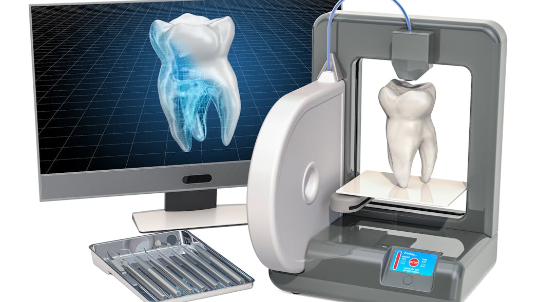
For almost 40 years,dentistry has been associated with some form of CAD/CAM technology. We have been scanning teeth and models for decades and using CNC technology to mill and manufacture restorations. This process has been purely reductive, meaning a block or ingot is ground or milled down to produce the final restoration. However, with the advent of additive manufacturing—think 3D printing—we are entering a new era of dental manufacturing, and it’s going to be a very exciting time.

Traditional milling of dental restorations began with diamond grinding of glass ceramics. Feldspathic blocks were ground using dual-motor CNC machines to create inlays/onlays and crowns. This process would sometimes take upward of 30–45 minutes. At the time, this was an incredible innovation, but today that amount of time would be simply unacceptable.

Fast-forward to today and the milling processes are light years ahead of where we started. Today, I can send a restoration from my CEREC Primescan, which was designed using only a few clicks, to my Primemill and have a full-contour zirconia restoration manufactured in less than five minutes. It’s incredible to think how much further we can go with the speed, accuracy, and efficiency we have in our current technology. And that is only in the chairside world.

Our laboratory partners have even more incredible technology that allows them not only to manufacture high-quality restorations efficiently, but on a much larger scale. Using large five-axis milling machines, laboratories can manufacture dozens of restorations at once out of a single puck of material. This has helped reduce the overhead for labs and increase their output. The modern dental lab technician may no longer have a CDT degree, but instead a computer science and graphic design background. Design and milling restorations will forever be a computer-guided process.

Within the last five to seven years, the world of 3D printing has exploded onto the dental scene. Formlabs was one of the first manufacturers to target the dental market with its Form2 printer. Using a vat filled with uncured resin, a very detailed laser would systematically cure the resin onto a build platform to create dental models from digital impressions. My first 3D printer would typically take 10–12 hours to manufacture a model, and that was completely acceptable at the time. In fact, it was exciting to finally eliminate alginate, stone, and a model trimmer!

Today, companies such as SprintRay and Dentsply Sirona are creating powerful and innovative 3D printing solutions that are speeding up the manufacturing process by leaps and bounds. On my Primeprint, I can produce a set of models in fewer than 30 minutes, which was unheard of just a few years ago.

With the rapid pace of innovation, research, and development—specifically regarding biocompatible materials and resins—it’s only a matter of time before additive manufacturing becomes the standard in dentistry. It took us almost 40 years to take a milled crown down from 40 minutes to five, and only two to three years to take a 12-hour printing process down to 30 minutes. We are already printing surgical guides and splints and provisionals today. I imagine within a few years, 3D printing of definitive restorations will have a significant place in the market. Regardless, I’m excited to just be along for the ride.  

Editor’s note: This article appeared in the May 2022 print edition of Dental Economics magazine. Dentists in North America are eligible for a complimentary print subscription.
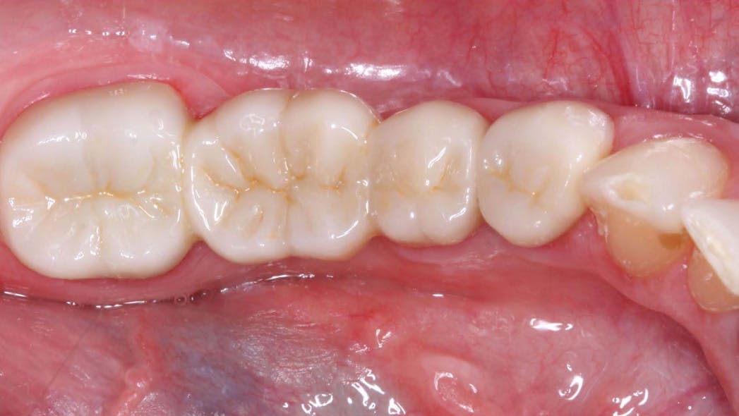
Which crown types are best for what situations?

Figure2

View Image Gallery 

Q: There is enormous confusion in the dental marketplace regarding which indirect restorations should be used and where in the mouth they should be used. The ads are not only confusing, but, in my opinion, they are also too optimistic. The significant commercial information claiming unprecedented success for specific restorations seems unrealistic. What is the state of the art for indirect restorations? What should be used and where in the mouth? How does heavy occlusion factor into the decision? Can optimum esthetics be achieved for all new materials? I would appreciate knowing what Clinical Research (CR) Foundation has found in long-term clinical studies.

A: I answered some of the same questions in “Which crown goes where?” but since then, many changes have occurred, more research is available, and questions keep coming up frequently. Importantly, the research on the various indirect restorations is starting to mature and provide some answers. In this article, I will give you a status report on the most-used crown types and their current success in CR/TRAC (Technologies in Restoratives and Caries Research Division of the CR Foundation) in vivo research. The information is divided into several locations in the mouth along with my suggestions as to the different strength and esthetic needs for those locations.


Also by Dr. Christensen:

Increasing practice activity

Are endodontic posts really necessary?


The following overall statements relate to my answers.

  • It is reported by large US dental labs that over 74% of indirect restorations are for single teeth.1
  • Large labs report that ceramic indirect restorations currently comprise over 90% of the total indirect restorations made. The majority of those indirect restorations are milled from one of the zirconia variations, some are milled or pressed lithium disilicate, and a small number are conventional porcelain-fused-to-metal (PFM), or polymer.1
  • A very conservative estimate is that about one-third of the adult population have either grinding or clenching bruxism, a highly important characteristic for restoration selection.
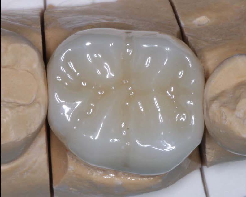
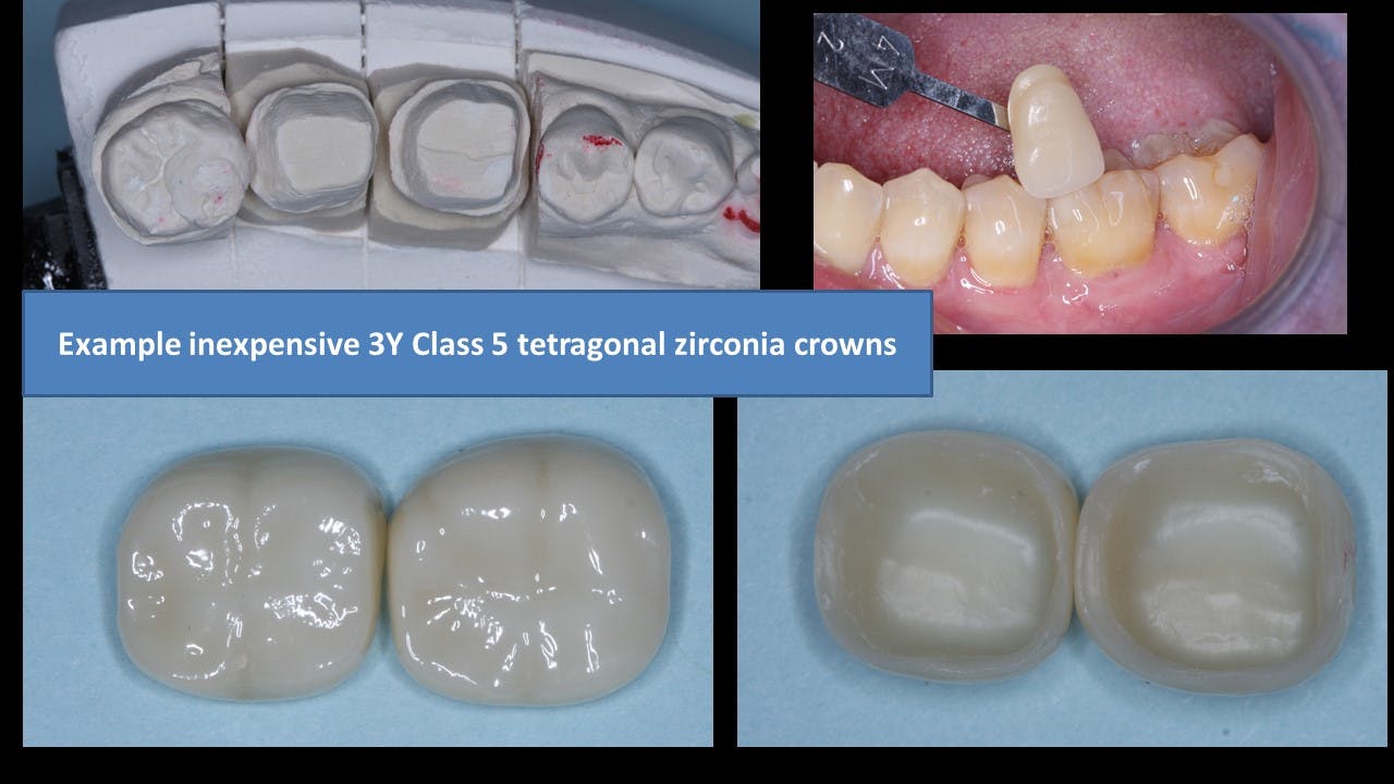
Figure 1: Class 5, tetragonal, 3Y zirconia—the original BruxZir has revolutionized the dental profession. It is now well proven to be the strongest and most durable ceramic restoration in dentistry, but color challenges still exist.

It can be concluded from the previous statements that different types of indirect restorations are present and that their various physical and esthetic characteristics relate to where they should be used. It is also important to note that additional long-term research is needed to confirm some of my suggestions.

Molars

Optimum strength is available by using class 5, 3Y, tetragonal zirconia (figure 1). This is the original BruxZir formulation, now available from many Glidewell laboratories. Similar products are produced by other companies under other names. It is often called LT (“low translucency”) zirconia by dentists and labs. The strength and durability of this zirconia category have been confirmed through 11 years of in vivo research by the TRAC Division.

Figure 2: A three-unit fixed prosthesis and a separate single crown milled from class 5 zirconia with a thin layer of veneering ceramic/glaze to make it an acceptable color. However, the thin veneer/glaze will eventually wear off the occlusal surface.

But, as most dentists know, this zirconia category has less-than-desirable esthetic qualities unless coated with layering ceramic or stained in the presintered zirconia stage (figures 2 and 3). Some dentists do not object to the unmodified color of this zirconia category for molars since it is not usually visible in the posterior of the mouth.

This zirconia formulation is well proven and has had unprecedented clinical success and lack of breakage. However, labs and manufacturers primarily promote the more esthetic forms of zirconia, identified as class 4 cubic zirconia. It is often described as HT (high translucency), or esthetic zirconia. This form of zirconia has lower strength than class 5 zirconia, and it still lacks long-term research for use in high-strength needs.

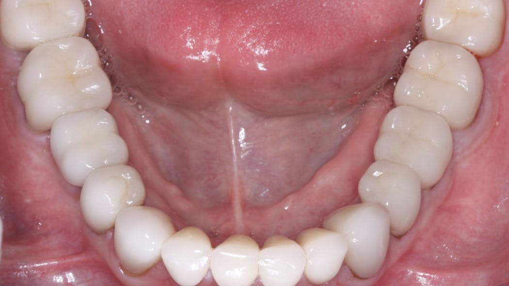
Figure 3: A four-unit fixed prosthesis milled from class 5 zirconia placed in a patient with extreme bruxing with pigment placed on the zirconia in the presintered stage to make the color acceptable.

Currently, clinical research has mixed results, indicating promise for class 4 zirconia formulations but also some potential challenges. Since this formulation has been available for only a few years, you and your peers are doing much of the observational clinical research on these materials in your practices. Long-term clinical research is still needed to validate the use of this zirconia form for molars, bruxing patients, long-span fixed prostheses, and other high-strength clinical needs.

Premolars

IPS e.max (lithium disilicate) is classified as a class 3 ceramic restoration. It is very well proven for single premolar restorations by both controlled studies and millions of such restorations placed internationally. As you know, it has unprecedented high esthetic qualities and strength (figures 4 and 5).

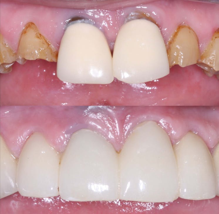
Figure 4: IPS e.max can cover even dark-colored teeth if it is at least 1.0 mm thick in all axial aspects. The left lateral incisor replacement is an implant. It has a thin metal opaqued coping placed over the implant, making the color of the class 3 ceramic restoration (lithium disilicate) acceptable.

For nonbruxers, IPS e.max can be used safely for single premolars and select three-unit fixed prostheses involving both premolars and anterior teeth. It is suggested that at least 1.0 mm of IPS e.max thickness is present on all axial walls and 1.5–2.0 mm thickness on the occlusal surface for optimal strength. However, more fractures have been reported on multiple-unit lithium disilicate fixed prostheses in the premolar to anterior area than on single teeth.

Should IPS e.max be used on premolars in bruxing patients? Some practitioners are using it in bruxing situations because of its great success in nonbruxers. The only alternatives are porcelain-fused-to-metal (PFM) or class 4 zirconia. Selecting an appropriate restoration for a bruxing patient requires that the dentist have personal knowledge of the patient, the anticipated occlusal loading, and 

Figure 5: The strength and esthetics of IPS e.max for single crowns is unexcelled by other materials.

any esthetic needs.

Should class 4 cubic zirconia restorations be used for premolars? These modified zirconia forms are being highly promoted for such situations. Clinical observation by CR evaluators has been promising, but those observations are only short term. Developing challenges have already been noted in some brands by CR’s in vitro microscopic research, indicating the use of class 4 zirconia with caution until additional long-term research is available.

At present, IPS e.max is a well-proven product for single premolars and select three-unit fixed prostheses involving premolars.

In situations involving high-strength needs, such as in bruxing patients, color-modified class 5, 3Y zirconia is still a more-proven concept. Additional clinical research will determine if class 4 zirconia will be adequate for patients who are bruxers.

Anterior teeth

Anterior teeth usually have the least need for strength. The statements on premolars apply directly to anterior teeth, but esthetic acceptability is more important in the anterior area of the mouth.

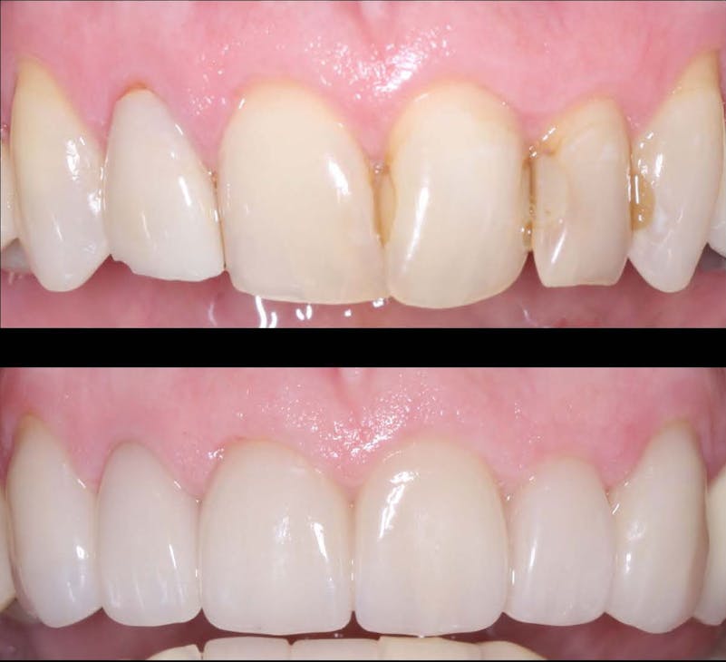
If the restoration is for single teeth and the patient is not a bruxer, IPS e.max currently is the optimum restoration.

If a three-unit (or larger) fixed prosthesis is needed, color-modified class 5, tetragonal, 3Y zirconia may be optimum, especially for a bruxing patient, but such zirconia requires an artist/technician to achieve the best esthetics.

Should class 4 cubic zirconia be considered for anterior three-unit (or larger) fixed prostheses? The same challenges are present as those for premolars. More long-term research is needed. How long? At least several years of research on many brands. That research is beginning to come forth, but there are still numerous questions. If using class 4 zirconia for anterior restorations, I suggest observing the restorations carefully with high-power loupes at each recall appointment. Look for pits, minor cracks, excessive wear on opposing teeth, or other maladies. These challenges have been seen on some class 4 zirconia brands in preliminary research. It is our hope that class 4 zirconia brands will soon prove themselves in clinical research. In the meantime, be observant and cautious.

Summary

Significant confusion is present about what type of indirect restoration is best for specific situations. Current evidence, both scientific and observational, support the use of class 5, tetragonal, 3Y zirconia. However, this formulation has esthetic challenges that must be overcome. Class 4 cubic-containing zirconia has many formulations. Many brands are currently proving themselves, but more years will be necessary for that proof to be solidified.

IPS e.max is well proven for near universal use in nonbruxers and limited use in bruxers. In the meantime, don’t forget the more than 120 years of success dentistry has had with cast-gold alloy and the more than 65 years of success with porcelain-fused-to-metal.

The immediate future appears to point to continued and expanded use of zirconia indirect restorations with a slow reduction in the use of the excellent, well-proven IPS e.max.  

Reference

  1. Based on data from Glidewell Labs.

Author’s note: The following educational materials from Practical Clinical Courses offer further resources on this topic for you and your staff.

One-hour videos:

  • Cementing Restorations—Proven and Successful (Item no. 1921)
  • Impressions Can Be Simple and Predictable (Item no. 1922)

Two-day hands-on courses in Utah:

  • Restorative Dentistry 1—Restorative/Esthetic/Preventive with Dr. Gordon Christensen
  • Faster, Easier, Higher Quality Dentistry with Dr. Gordon Christensen

For more information, visit pccdental.com or contact Practical Clinical Courses at (800) 223-6569.

Editor’s note: This article originally appeared in the February 2022 print edition of Dental Economics.

Fracture Resistance and Fracture Behaviour of Monolithic Multi-Layered Translucent Zirconia Fixed Dental Prostheses with Different Placing Strategies of Connector: An in vitro Study

Purpose: To evaluate the effect of different placing strategies performed in the connector area on fracture resistance and fracture behaviour of monolithic multi-layered translucent zirconia fixed dental prostheses (FDPs).
Materials and Methods: Thirty 3-unit monolithic FDPs were produced and divided into three groups (n = 10) based on the different strategies for placing the connector area of FDPs in multi-layered zirconia blank with varying contents of yttria ranging from 4 to 5 mol%. The groups were as follows: FDPs with connectors placed in dentin layer with 4 mol% yttria content, FDPs with connectors placed in gradient layer, and FDPs with connectors placed in translucent layer with 5 mol% yttria content. A final group (n = 10) of conventional monolithic zirconia with a monolayer of yttria content (4 mol%) has been used as a control group. The specimens were artificially aged using thermocycling and pre-loading procedures and subsequently loaded to fracture using a universal testing machine. Fracture loads and fracture behaviour were analyzed using one-way ANOVA and Fisher’s exact tests and statistically evaluated (p ≤ 0.05).
Results: There were no significant differences in fracture loads among the groups based on the placing strategies of the connector area of the FDPs in the multi-layered translucent zirconia blank (p > 0.05). There was no significant difference in fracture loads between monolithic multi-layered translucent zirconia and conventional monolithic translucent zirconia materials (p > 0.05). Fracture behaviour of FDPs with connector area placed in translucent layer differed significantly compared to FDPs with connector area placed in dentin layer and FDPs in control group (p = 0.004).
Conclusion: The placing strategies of the connector used in the computer aided design and manufacturing procedures do not considerably affect fracture resistance of monolithic FDPs made of multi-layered translucent zirconia. Monolithic FDPs made of multi-layered translucent zirconia show comparable strength to FDPs made of conventional translucent zirconia, but with different fracture behaviour.

Keywords: all-ceramic restorations, computer-aided design\manufacturing, fracture load, multi-layered zirconia, Y-TZP

Introduction

Yttria-stabilized tetragonal zirconia polycrystal (Y-TZP) is the most commonly used oxide ceramic material in Restorative Dentistry. This is related to its superior fracture strength and unique toughening properties.1,2 However, owing to its poor optical properties, Y-TZP based restorations must be veneered with translucent glass-ceramic materials in many clinical situations. Although the high success rate of veneered Y-TZP restorations has been reported to be over 90%, clinical complications such as veneer chipping and connector fracture still occur.3–5 Moreover, the use of veneered Y-TZP restorations requires removing more underlying tooth substance to provide enough space for the material. For those reasons, there is a general preference for shifting toward monolithic Y-TZP restorations, with challenges in achieving esthetical requirements without compromising the overall strength.6–8

The main drawback of using Y-TZP material as a monolithic restoration is the low translucency, resulting in poor esthetics.6,9–11 Scattering of light in Y-TZP and subsequent reduction of light transmittance mainly occurs at grain boundaries, pores, and secondary phases.6,9–11 However, enhanced optical properties of this material have been achieved by modifying the microstructure, for example, through altering the yttria (Y2O3) content and applying different sintering conditions.12,13 Shorter sintering times result in smaller grain size and thus an increase of the light transmittance of the final dental zirconia.12 Furthermore, it has been shown that the change of dopant contents, such as lanthanum oxide and aluminum oxide, improved the optical properties of zirconia.14 From a material point of view, the mechanical properties of Y-TZP are negatively affected by enhancing the translucent properties of the material.15,16 The more translucent the zirconia is, the lower the fracture strength.15,16

Recently, a new multi-layered translucent zirconia material, with a natural progression of shade and translucency, has emerged in the dental market to mimic natural teeth closely. This material is indicated to produce monolithic restorations in both the anterior and posterior regions. There are two types of multi-layered translucent zirconia on the market: 1) Multi-layered zirconia with different colour saturations in the different layers but the same yttria content throughout all layers, and 2) Multi-layered zirconia with different translucency in the different layers as a result of varying yttria contents in the different layers. Thus, the strength and toughness of the layers with different yttria contents are expected to be different. During computer-aided design and manufacturing (CAD/CAM) procedures, dental technicians can use different placing strategies to place the fixed dental prosthesis (FDP) in multi-layered translucent zirconia blank before milling. Previous studies showed that the main fracture origin leading to the failure of the prostheses is located at the gingival side of the connector area, which is linked to the development of stress concentrations in the connector when different loads are applied to the FDPs.17,18 Accordingly, in practice, the fracture resistance of the FDP, especially in the connector area, might be affected depending on how the placing strategy has been performed by the dental technician during CAD/CAM procedures. It is not known, however, if the different placing strategies of the connector, during computer manufacturing of zirconia blanks, might affect the fracture resistance of the final restoration made of the new multi-layered translucent zirconia material, since the strength varies between the different layers of zirconia.

Therefore, the present study aimed to evaluate the effect of the different placing strategies performed in the connector area on fracture resistance and fracture behaviour of monolithic FDPs made of multi-layered translucent zirconia. The null hypothesis is that there is no difference in fracture resistance and fracture behaviour of the FDPs made of multi-layered translucent zirconia based on the placing strategies performed in the connector area.

Materials and Methods

Study Design

Thirty 3-unit monolithic FDPs were produced and divided into three groups (n=10) according to the different strategies for placing the connector area of the FDPs in the multi-layered translucent zirconia blank (IPS e.max ZirCAD MT Multi, Ivoclar Vivadent, Schaan, Liechtenstein) (Figure 1). The groups were as follows: FDPs produced with the connectors placed in the dentin layer with 4 mol% yttria, FDPs produced with the connectors placed in the gradient layer, and FDPs produced with the connectors placed in the translucent layer with 5 mol% yttria. A final group (n=10) of conventional monolithic zirconia with monolayer of 4 mol% yttria content has been used as a control group (IPS e.max ZirCAD, Ivoclar Vivadent, Schaan, Liechtenstein). The FDPs were cemented using compatible resin cement onto abutment models made of a polymer material (POM C glass infiltrated). The specimens were artificially aged using both thermocycling and cyclic fatigue procedures before they were loaded to fracture. Fracture loads and fracture behaviour were subsequently analyzed and evaluated statistically p ≤0.05.

Figure 1 Illustrations show different placing strategies of the connector area of the FDPs in multi-layered zirconia blank through computer-aided manufacturing software. The double-headed black arrow represents moving the FDP in translucent layer (5Y-TZP), gradient layer, and dentin layer (4Y-TZP) of the multi-layered zirconia blank before milling.

Specimen Preparation

For the preparation of the teeth, a plastic model of a mandibular jaw was used (KaVo YZ; KaVo Dental GmbH, Biberach, Germany). The preparations were made on the canine (43) and premolar (45) and were designed to provide space for Y-TZP material with a 120° chamfer and 15° convergence angle. The teeth preparations were conducted by prosthodontist. After the preparations were conducted, a full-arch impression using silicone material (President; Coltene AG, Altstätten, Switzerland) was made and poured with die stone material (Vel-Mix; Kerr Corp, Orange, CA). A master cast was produced from the die stone, and subsequently, a wax-up (1.5–3 mm) of the FDP was made by professional dental technician. The wax-up was scanned with a double-scan technique using a dental laboratory scanner (D900L; 3Shape, Copenhagen, Denmark). Data from the scanner were transferred to a computer loaded with computer-aided design (CAD) software. The design of the FDP connector was a round shape and the dimensions for all the FDP connectors were adjusted to 3 mm x 3 mm. The occlusal thickness of the retainer core was set to 1 mm, and the axial wall thickness was set to 0.8 mm with a 0.5 mm cervical margin. After the adjustments, the CAD file was sent to a milling center (Cosmodent AB, Malmö, Sweden) to produce the FDPs. The same sintering protocol for the two zirconia materials has been used following the manufacturer instructions. The CAD file was used to produce the abutment models made from a polymer material (POM-C GF25; Mekaniska AB, Simrishamn, Sweden) with a modulus of elasticity comparably close to dentin (9 GPa).

Artificial Aging, Cementation, and Load to Fracture Test

All FDPs were subjected to artificial aging, beginning with thermocycling. In a thermocycling device (THE-1100; SD Mechatronik GmbH, Feldkirchen-Westerham, Germany) containing two water baths, the FDPs underwent 10,000 thermocycles at two different temperatures, 5 and 55°C. Each cycle lasted for 60 seconds, 20 seconds in each bath and 20 seconds to complete the transfer between the baths.4,19–22 The cementation of the FDPs to the abutment models was completed using a dual-polymerized resin cement (Panavia V5; Kuraray Medical Inc., Okayama, Japan) according to the manufacturer’s recommendations. However, before cementation, the abutment models were air-abraded with 50 µm aluminum oxide using an air abrasion device (Basic Quattro IS; Renfert GmbH, Hilzingen, Germany) as well as treated with two primers (Tooth Primer, Clearfil and Ceramic Primer; Kuraray Medical Inc) following the manufacturers’ instructions. The FDPs were cemented to the abutment models with a standardized seating load of 15 N in the direction of insertion. A calibrated curing lamp (Heraeus Translux® Power Blue®, Heraeus Kulzer GmbH) was used according to the manufacturer’s recommendations to initiate the curing. Ultimately, excess cement was removed with a scalpel (AESCULAP® no. 12, Aesculap AG & Co, Tuttlingen, Germany). The specimens were stored in a humid environment at a temperature of 37°C before cyclic fatigue. The last step of artificial aging was cyclic fatigue using a pre-loading machine (MTI Engineering AB; Lund, Sweden/Pamaco AB, Malmö, Sweden). The cemented FDPs were submerged in distilled water at 10° of inclination towards the tooth axis and went through 10,000 cycles of 30–300 N at a frequency of 1 Hz. A 4 mm stainless ball was placed on the occlusal surface of the connector area between teeth 45 and 44 of the bridges to apply mechanical cyclic loads.4,19–22

After artificial aging, all FDPs were installed in a test jig at 10° inclination towards the axial direction using a universal testing machine (Instron 4465, Instron Co. Ltd, Norwood, MA, USA), (Figure 2) as was suggested in previous laboratory studies.4,19–22 The load was applied on the pontic using a specialized stainless-steel intender. Throughout loading, all the FDPs were submerged in water. The crosshead speed was set at 0.255 mm/min, and the fracture was defined as follows: visible crack, load drop or an acoustic event, whatever occurred first.4,19–22 The load at fracture was then registered.

Figure 2 Illustration of the specimen in a test jig at 10° inclination in cyclic fatigue and load to fracture tests. All specimens were submerged in water during the tests.

Fracture Behaviour Analysis

The fracture surfaces of the FDPs were analyzed by two examiners. A gross visual and microscopic assessments (Leica DFC 420, Leica Application Suite v. 3.3.1, Leica Microsystems CMS GmbH, Wetzlar, Germany) were performed to classify fracture behaviour according to the location of fracture into: fracture at the distal connector, fracture at the mesial connector, complete fracture of the FDP (involving fracture of the retainer).

Statistical Analysis

The differences in fracture resistance among the groups were analyzed using one-way ANOVA, followed by Tukey’s post hoc test (IBM SPSS Statistics 25). The differences in fracture behaviour among the groups were analyzed using Fisher’s exact test. The level of significance was set to p ≤0.05. The statistical analysis was performed by an experienced professional statistician. Power analysis was based on previous studies where differences regarding the level of significance and standard deviation were detected among the zirconia-based specimens.17,19–21

Results

Loads at fracture, levels of significance, fracture behaviour for all groups are summarized in Tables 1 and 2. There were no significant differences in fracture loads among the groups based on the different strategies for placing the connector area of the FDPs in the multi-layered zirconia blank (p >0.05). There was no significant difference (p >0.05) in fracture loads between the two different materials: monolithic multi-layered translucent zirconia and conventional monolithic translucent zirconia materials.

Table 1 Load at Fracture in Newton (N)

Table 2 Distribution of Fracture Behaviour

Three types of fracture behaviour were registered after load to fracture test: fracture at the mesial connector propagating through the pontic, fracture at the distal connector propagating through the pontic, and complete fracture involving the retainer (Figure 3). Fracture behaviour of the FDPs with connector area placed in the translucent layer (5Y-TZP) differed significantly compared to the FDPs with connector area placed in the dentin layer (4Y-TZP) and the FDPs in the control group (p ≤0.05).

Figure 3 Different types of fracture behaviour. (A) Fracture at distal connector; (B) complete fracture; (C) fracture at mesial connector.

Discussion

The null hypothesis of this study was rejected since fracture resistance of the FDPs showed no significant differences among the groups based on the different placing strategies performed in the connector area during computer manufacturing of the FDPs. However, the results showed that the different placing strategies performed in the connector area affect fracture behaviour of the three-unit FDPs.

One of the common methods to improve the translucency of dental zirconia is by changing the amount of yttria content, which results in a greater portion of the optically isotropic cubic phase without light scattering at the grain boundaries.14,15The major phenomena related with the enhanced translucency of polycrystalline zirconia-based ceramics is the reduction of birefringence, the light scattering promoted by a material with anisotropic refractive index. Tetragonal zirconia phase is birefringent, however, by increasing yttria content the precipitation of cubic zirconia, which is isotropic and do not experience birefringence, is favoured and an enhancement of the transmitted light fraction is experienced.23–25 This, on the other hand, compromises the strength and toughness of the cubic zirconia because it does not undergo stress-induced transformation.14,23–25 In the present study, the FDPs made of multi-layered translucent zirconia were divided into three groups: dentin, gradient, and translucent, based on the content of yttria ranging from 4 to 5 mol%. The groups with the connectors placed in the gradient and the translucent layers presented higher standard deviation values than the dentin and control groups. This might be explained by the fact that the gradient layer combines different microstructures of both the translucent and the dentin layers, which results in varying mechanical properties. Thus, the FDPs with the connectors placed in the layer consisting of a microstructure primarily composed of dentin (4Y-TZP) withstand higher fracture loads. The opposite applies to the FDPs with the connectors placed in the layer consisting of a mainly translucent microstructure, namely 5Y-TZP. These findings are in line with previous studies, which concluded that translucency affects the mechanical properties of zirconia.15,23–25 Although the differences of the results were not statistically significant, the numerical differences among the groups in this study, together with the findings of previous studies, confirm the effect of enhanced translucency on the mechanical properties of Y-TZP. Moreover, it is noteworthy that the limitations of the methodology used in this study might have influenced the results. For geometric reasons, it is impossible to place the whole reconstruction in one layer in the multi-layered translucent zirconia blank without infringing the minimum dimensional demands of the FDP. This means that the critical part of the connector area, the gingival portion, where the highest stress concentrations occur during loading, will probably not be entirely located in solely one layer.17,18 This technical limitation means that study findings need to be interpreted cautiously.

Many studies have investigated the adverse effects of the other methods of enhancing the optical properties of zirconia on mechanical properties. For instance, although doping of metal oxides improves the optical properties of zirconia, this may affect adversely the mechanical and biological (cytotoxic) properties of zirconia.14 Other fabrication techniques such as colouring of pre-sintered zirconia for enhancing the optical properties might be necessary in many clinical cases. Previous studies have shown the effect of such colouring techniques on the mechanical and optical properties.26,27 Nevertheless, a very recent study investigating new multi-layered translucent zirconia material showed no differences in neither microstructure nor translucency between the different layers.28 Only colour pigment composition is different between the layers within each multi-layered translucent zirconia blank. The same study revealed that lanthanum oxide doping improved the translucency without diminishing the mechanical properties of the multi-layered translucent zirconia, which is the main goal when developing high esthetical monolithic dental zirconia.

Considering fracture behaviour, this study showed that most fractures started from the connector area (mesial or distal) and propagated through the pontic during loading. This is in agreement with previous studies, which concluded that critical tensile stresses mostly develop in the gingival embrasure of the connector, result in failure of prosthesis.17,18 However, there were significantly more complete fractures (involving the retainer) in the FDPs with connector area placed in the translucent layer (5Y-TZP) compared to the FDPs of the other groups. This finding could be expected theoretically since the translucent layer has a microstructure that is less resistant to fractures, as previous studies have shown.15,23–25 It is noteworthy that fracture behaviour analysis in this study aimed to show the fracture initiation and propagation pattern under a light microscope and evaluate the ability of the test to mimic the clinical failures of dental restorations. Sophisticated fractographic analysis using a scanning electron microscope, however, might provide more details on fracture behaviour.

When conducting an in vitro study to evaluate the mechanical properties of new material, a laboratory setup simulating the oral environment and the complex forces of mastication is of great importance. One of the limitations of in vitro studies is the difficulty to choose which aging procedures would produce comparable clinical results. Previous studies have investigated the effect of artificial aging procedures, that used to mimic the clinical situation, on the longevity of ceramics. Despite that some of those studies fail to show a direct relationship between aging procedures and fracture resistance of ceramics,29 most agree that they have a significant effect on the longevity of ceramic materials.30–32 Therefore, there is no consensus regarding the effectiveness of aging tests or a specific aging protocol, but it was reasonable, however, to perform such procedures in the present study to allow for comparison of the results of other studies carried out by the same research group with this specific protocol.4,19–21 The FDPs were mounted with a 10 of inclination relative to the load direction in the load to fracture test. This angle of inclination has been used in many previous studies and was initially suggested by Yoshinari and Derand.4,19–22 However, the mechanical load to fracture test performed in a laboratory study can never completely reproduce loads and environmental influences as in the clinical situation but can still give important information. Furthermore, to obtain realistic fracture load values and compare these values with previous studies, replicating the real clinical situation concerning mechanical support is crucial.33 Therefore, all FDPs were cemented onto abutment models made of a material with a modulus of elasticity close to dentin. The cementation procedure was performed according to the manufacturer’s recommendations, and the same cement was used for all groups. Since in vitro studies have shown that thermocycling affects the bond strength of cements, all FDPs were cemented after this stage to avoid partly loose prostheses at the subsequent cyclic fatigue and load to fracture tests.31,32

Since adequate communication between the dentist and the dental technician is essential for successful dental restorations, it is a prerequisite for dentists to gain knowledge of the dental material that is required. This study has shown that the different strategies for placing the FDP in the blank during the CAD/CAM process do not have a critical effect on the mechanical properties of the translucent multi-layered zirconia FDPs. Thus, this facilitates the process of ordering for the dentist who does not have to pay regard to the technical aspects. In vitro studies, in line with the present one, are of great importance to evaluate new dental materials before using them in a clinical situation, thus safeguarding patient safety.

Conclusion

Within the limitations of this laboratory study, the following conclusions can be drawn: the placing strategies of the connector used in the computer aided design and manufacturing procedures do not considerably affect fracture resistance of monolithic FDPs made of multi-layered translucent zirconia. Monolithic FDPs made of multi-layered translucent zirconia show comparable strength to FDPs made of conventional translucent zirconia, but with different fracture behaviour.

Improving the color match of zirconia crowns

2109 De Cchr P03 610066ead811f


QUESTION:

Most of the zirconia crowns and fixed prostheses coming from my labs lately are too light in color, despite my providing preoperative photos and descriptions to the technicians. I have tried several different laboratories, and I am still frustrated with the mismatched colors. Although zirconia crowns are strong, I believe their color-matching is severely lacking. My colleagues have expressed similar color mismatch as well. What can be done to improve this obvious problem?

ANSWER:

I agree completely with your criticism of the current generation of zirconia crowns. The Technologies in Restoratives and Caries Research (TRAC) Division of the Clinical Research Foundation has accomplished long-term in vivo research on zirconia crowns and found their strength to be fantastic and their service over many years to be outstanding. However, only recently has the color of zirconia crowns been improved so they are more acceptable.

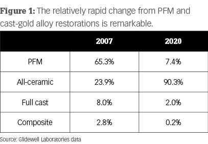
The changes to improve color and translucence of the crowns has been successful, but it also has had some negative effects. The information in Figure 1 is from Glidewell Laboratories and shows remarkable changes over the last several years as ceramic restorations have become mainstream. What are manufacturers doing to improve the esthetics of zirconia restorations? To answer that question, let’s discuss the various types of zirconia.

Zirconia and its various forms

When observing the 2020 data from Glidewell Laboratories in Figure 1, it is obvious that zirconia wins the popularity contest. As you probably know, Glidewell started this clinical revolution with BruxZir, which soon dominated the market. Zirconia-based crowns have been available for more than 21 years, and full-zirconia crowns have been available for about 10 years.

2109 Dec57 60 Chr

In the first few production years, the original zirconia-based crowns had challenges, but since then those problems have largely been overcome. Despite that, the use of zirconia-based restorations has not grown markedly.

Full-zirconia restorations of various formulations are extremely popular and state-of-the-art. But zirconia is not zirconia. You are undoubtedly familiar with the ADA/ISO classification of ceramic crowns, which we previously published in the July 2019 issue of Clinicians Report. (Visit cliniciansreport.orgto read the article in detail.)

The initial BruxZir formulation was classified as class 5 tetragonal zirconia. This is probably what you will receive if you order class 5 or 3Y (3 molar percent yttrium oxide) tetragonal zirconia. This formulation has been used in nondental applications in various industries for more than 30 years. It is the strongest form of zirconia and has optimal transformation toughening (lack of crack propagation) during service. Unfortunately, this is the iteration of dental zirconia most difficult to make esthetically acceptable.

If you are planning a long-span fixed partial denture (FPD) or treating a patient with bruxism, this is an excellent choice shown to be comparable in strength to porcelain-fused-to-metal (PFM) in our long-term TRAC studies. It can be made esthetically acceptable for some situations by presinter staining or optimum use of external stains.

Manufacturers have been experimenting with making 3Y zirconia more esthetic. The most successful and easiest method to date is to increase the translucence by adding more translucent material to the zirconia. ADA/ISO classifies this type of zirconia as class 4 cubic-containing zirconia. It is also identified as 4Y or 5Y or combinations of those percentages. Although additional long-term clinical research is still needed for the so-called esthetic zirconia, class 4 zirconia currently appears to be adequate for singles, short-span FPDs, and other situations not requiring the strength of 3Y zirconia.

Why do you need to know the zirconia classifications?

Your knowledge of these classifications allows you to communicate with salespeople and laboratory technicians, as well as understand what materials you are putting into your patients’ mouths. Knowing this, you’ll better understand the changes that are being made in the original dental zirconia restorations.

Adding more translucent materials to the zirconia. Additional oxides are now being added to the original Glidewell formulation of zirconia (3Y, class 5 zirconia versions) by many manufacturers. This formulation change improves translucency and esthetics. However, the change to 4Y or 5Y or combinations of those reduces the overall strength of zirconia and its ability to “heal” microcracks. (Higher transformation toughening increases the lack of subsequent crack propagation.) Although this procedure reduces strength, clinical success is currently being proven and reported in the field by practicing dentists. The esthetic characteristics of some of these class 4 restorations can rival IPS e.max (Ivoclar Vivadent).

Figure 2: External staining and glazing of zirconia is commonly accomplished in dentistry. Unfortunately, it has some negative characteristics over the long term.

Placing relatively thick layers of stain and glaze on the external of the restorations. Many labs are firing ceramic on the outside of the mismatched zirconia to improve the zirconia color (figure 2). In our research, these additions are usually feldspathic ceramic and are showing wear on the opposing teeth. There is also a slow but continuing loss of the added superficial layers. If the external layers are thin, the result is a slow change back to the original zirconia colors.

Internal staining of zirconia. Some labs are staining the 3Y, class 5 zirconia in the presintered stage, which improves the color significantly. However, this requires artistic technicians, more time, and a greater cost to the dentist (figure 3).

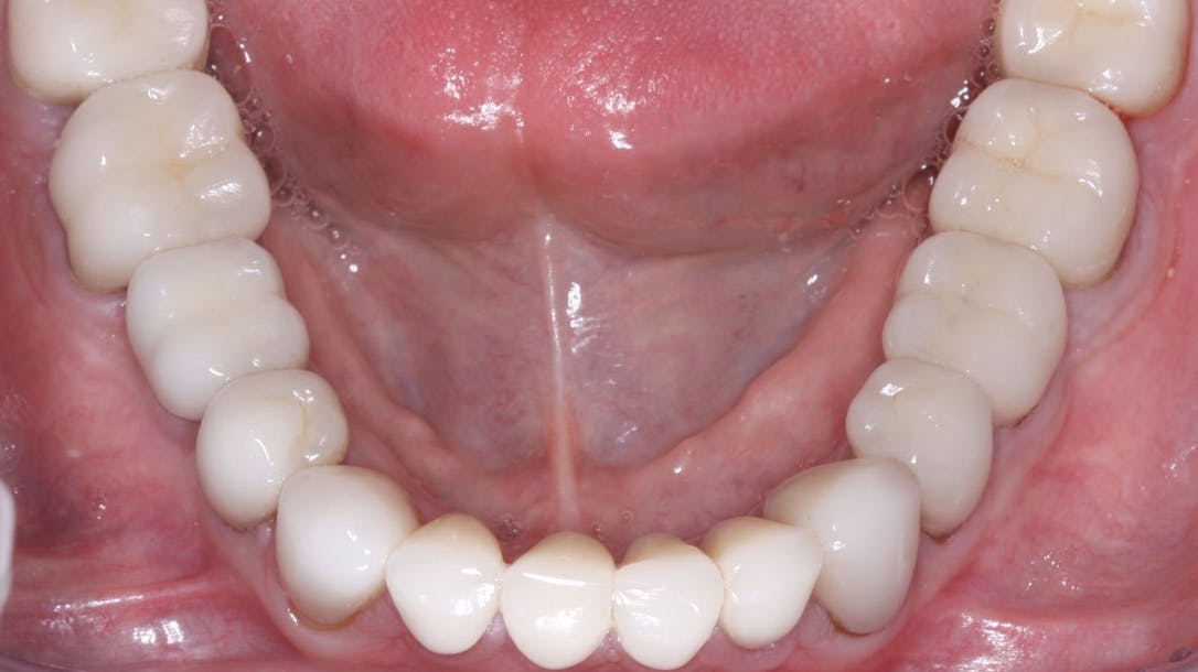
Figure 3: This rehabilitation, now in service for about five years, has IPS e.max on all teeth except the four molars. The 3Y, class 5 tetragonal full-strength zirconia on the molars was stained in the presintered stage, resulting in esthetics similar to e.max but without external staining.

Currently, most indirect restorations are ceramic, and that trend will undoubtedly continue to grow. Metal and porcelain-to-metal restorations are showing a relatively rapid decrease in use in dentistry.

Is there a dental laboratory problem?

Yes! The laboratory industry has changed rapidly and significantly. Until recently, porcelain-fused-to-metal restorations were the norm. That situation then changed to nearly all ceramic restorations, with zirconia restorations being rapidly produced by milling machines that use computer-driven software and by lithium disilicate being pressed or milled.

Many artistic laboratory technicians are being replaced with highly competent, computer-savvy technicians who produce restorations at record speeds considered to be impossible by older technicians. As a result, restorations are less expensive but often less esthetically acceptable.

If needed, clinicians must seek artistically oriented laboratory technicians to accomplish the complex esthetic results desired for some patients, and consequently, we must expect to pay higher fees for these restorations. Esthetic restorations are available—certainly with lithium disilicate (IPS e.max) and, with effort, even zirconia.

Which indirect restorations are desirable and for what situations?

I have heard some dentists comment with mixed opinions on the esthetic dilemma about which you asked. Most dentists admit, to an embarrassing degree, that many zirconia restorations do not adequately match adjacent teeth, but they further comment that zirconia restorations in the posterior nonesthetic regions are far more esthetically acceptable than gold alloy or porcelain-fused-to-metal when the glaze and staining have worn off.

Porcelain-fused-to-metal

Don’t forget the 70-year success of these restorations, especially when patients have had proven success with previous PFM restorations in their mouths. The advantages and disadvantages of these restorations are well known. Many labs can make highly esthetic PFM restorations. They are especially useful in clinical situations requiring precision or semiprecision attachments, which are not currently available for zirconia or lithium disilicate restorations. An additional continuing use for PFM is long-span fixed prostheses. Although not as commonly used as in the past because of the availability of implants, PFM restorations are still occasionally needed.

Lithium disilicate (most common brand name IPS e.max)

Can you name any other proven successful type of indirect restoration that equals the superior esthetic result of e.max for single-tooth restorations? That question is easy to answer. Many crown types have been tried and were initially successful, but they failed the need for longevity. The success of e.max in selected three-unit anterior FPDs has been shown, but I suggest that the newer generations of zirconia (class 4 zirconia) will probably prove to be more successful for long-term clinical success in these anterior situations.

Summary

The crown revolution has changed almost every treatment plan that requires crowns or fixed prostheses. This article describes the state-of-the-art and makes suggestions about which materials to use, their advantages and limitations, as well as how to produce the esthetic result necessary for specific clinical situations.


Author’s note: The following educational materials from Practical Clinical Courses offer further resources on this topic for you and your staff.

COVID-19 Vaccines: 7 Things Your Dentist Wants You to Know

Mouth Healthy TM
A vial of COVID-19 vaccine

What do flossing, fluoride and the COVID-19 vaccine have in common? Preventing disease.

Your dentist cares for your mouth because your oral health is essential to your overall health. Throughout the COVID-19 pandemic, your dentist has been working to put your health and safety first by taking extra steps to prevent the spread of COVID-19 in the dental office

Now, we have COVID-19 vaccines to add to the other tools we’ve all been using to fight the pandemic — like wearing masks, washing our hands and avoiding crowds. Here’s what the CDC (and your dentist!) want you to know about COVID-19 vaccines.

1. The Vaccines are Safe and Effective

As doctors of oral health, credible scientific information is important to us when recommending treatments for our patients. While these vaccines were developed in a shorter time frame than some other vaccines, it’s important to know that the science behind them was not rushed. And by mid-November, 193 million people in the US were fully vaccinated.

As an additional safety measure, the CDC has set up expanded safety monitoring systems like the V-Safe smartphone tool to monitor vaccinations in real time. These systems have shown that serious side effects are rare. 

2. The Vaccine Won’t Make You Sick, But It Does Have Some Side Effects

There is no possible way COVID-19 vaccines can give you COVID-19. They might, however, come with some side effects that make you feel uncomfortable for a short time.

Because vaccines teach your body how to recognize and fight off a COVID-19 infection, you might feel some of the symptoms you’d get if your body were fighting off the real virus, such as a fever, according to the CDC. While unpleasant, this is actually a sign the vaccine is working in your body.

3. You Should Still Get the Vaccine Even If You’ve Had COVID-19

Those who have recovered from COVID-19 have some natural immunity that may protect them from getting sick again. Data from the CDC shows that vaccination of people who have had COVID-19 significantly improves their level of protection against being infected again and against having serious COVID-19 illness. The CDC recommends that people who’ve had COVID-19 still get the vaccine.

4. Get All Recommended Doses

Generally, if you are receiving the Pfizer or Moderna vaccines, you need two doses to get the same level of efficacy seen in the clinical trials. For the Pfizer vaccine, the second dose is recommended three weeks after the first. For the Moderna vaccine, the second dose is recommended four weeks after the first. And if you get the Johnson & Johnson vaccine, you only need a single dose.

Everyone 16 and older can get a booster shot. If you received Moderna or Pfizer, you can get a booster six months after your second dose. If you received Johnson & Johnson, you are eligible for a booster two months after their first dose. 

You can also choose to “mix or match” your booster vaccine. The CDC’s recommendations allow for people to choose whether to receive the same vaccine as their initial doses or a different booster type. Visit the CDC’s website for the most up-to-date guidance around boosters or talk to your healthcare provider.

5. Vaccine Eligibility Expands to All Americans 5 and Older 

Anyone 5 and older can now receive a COVID-19 vaccine. Children and adolescents 5 – 17 are eligible to receive the Pfizer vaccine, and anyone 18 or older may receive any of the currently available vaccines. 
The CDC created easy-to-use tools to help you find a vaccine nearby:

  • Visit Vaccines.gov to search by vaccine type and zip code.
  • Text GETVAX (438829) for English or VACUNA (822862) for Spanish to receive vaccine sites on your phone.
  • Call the National COVID-19 Vaccination Assistance Hotline at 1-800-232-0233.

6. Mask Guidance: Depends on the Environment

Vaccines can help you resume the activities you enjoyed doing most before the pandemic. Being vaccinated is your best protection against becoming seriously ill with COVID-19. However, with the rise of the more infectious Delta and now the Omicron variant, the CDC has updated its guidance to recommend that everyone – regardless of vaccination status – wear masks in public indoor settings and even outside in areas with crowds. This is because these variants are very effective at infecting people who aren’t vaccinated, including those too young to be vaccinated. These variants can even infect some people who have been vaccinated and, even though the vaccine prevents them from getting seriously ill, they can spread the virus to others.

7. You Can Get the Vaccine If You Are Planning to Get Pregnant

The American College of Obstetrics and Gynecology (ACOG) recommends vaccination for those who are pregnant. Whether you are planning to get pregnant soon or in the future, you should still get the vaccine when it is available to you. The CDC states there is no evidence that the antibodies created from COVID-19 vaccines will cause problems with a pregnancy. The CDC also says there is no evidence that fertility issues are a side effect of the COVID-19 vaccine, or any other vaccine.

Have more questions? Talk to your dentist or physician. You can also visit the CDC’s website for more information about COVID-19 vaccines and find contact information for your local health department.https://findadentist.ada.org/search-widget?background=ffffff

Zircon Lab with Border_White

Choose The Right Dental Lab, The First Time

    Zircon Lab, LLC specializes in high end Zirconia (Bruxer), Aesthetic High Translucent Full Zirconia Crowns for Anteriors, e.max, and implants. All of our restorations are 100% made in the USA and we are the only dental lab to have an unconditional, 100% lifetime warranty.

Contact Info

8060 Reeder St. | Overland Park, KS 66214

+1 888.880.3383
contact@zirconlab.com

Monday - Friday: 8:00 am - 5:00 pm CST
Saturday & Sunday: Closed

Copyright 2023 Zircon Lab, LLC ©  All Rights Reserved