Thank You For Your File Submission
Someone from our team will be in touch with you soon!
News & Company Updates
Advancements in Dental Crown Materials: Enhancing Smiles with Innovation
The field of dentistry continues to witness remarkable advancements, and one area that has seen significant progress is dental crown materials. Gone are the days when traditional materials like metal or porcelain fused to metal were the only options available. Today, dentists can offer patients an array of cutting-edge materials, such as zirconia, lithium disilicate, and resin-based composites. These innovative materials bring numerous advantages, both aesthetically and functionally, revolutionizing the way dental crowns are designed and fabricated.
Zirconia, a type of ceramic, has gained popularity due to its exceptional strength, durability, and natural-looking appearance. It offers superior biocompatibility and is resistant to chipping, making it an ideal choice for patients seeking long-lasting and aesthetically pleasing dental crowns. Similarly, lithium disilicate, a glass-ceramic material, provides excellent translucency and can be precisely matched to the patient’s natural tooth color, resulting in highly esthetic restorations. Additionally, resin-based composites have evolved to offer improved strength, wear resistance, and lifelike esthetics, making them a versatile choice for anterior and posterior dental crowns.
The introduction of these advanced dental crown materials has revolutionized the dental industry, allowing dentists to provide patients with restorations that not only restore function but also blend seamlessly with their natural teeth. Patients benefit from enhanced durability, improved comfort, and superior esthetics, leading to increased satisfaction with their dental treatments. As the field of dentistry continues to evolve, dentists must stay informed about these advancements in dental crown materials to offer their patients the best possible care, ultimately transforming smiles and improving overall oral health.
In conclusion, advancements in dental crown materials have opened up a world of possibilities for dentists and patients alike. The introduction of zirconia, lithium disilicate, and resin-based composites has revolutionized dental crown treatments, providing superior strength, durability, and esthetics compared to traditional materials. As dentistry continues to progress, staying up-to-date with these innovative materials allows dentists to offer their patients the highest level of care, creating smiles that are both functional and aesthetically pleasing.
Tips for Choosing a Dental Lab
- Look for a dental lab with a good reputation: Do your research and look for a lab that has a good reputation in the industry. Ask for references and check online reviews to get a sense of the lab’s quality and customer service.
- Consider the lab’s certifications and accreditation: Look for a lab that is certified by organizations like the National Association of Dental Laboratories (NADL) or the American Dental Association (ADA). These certifications indicate that the lab meets certain standards of quality and professionalism.
- Consider the lab’s experience and expertise: Look for a lab that has experience and expertise in the specific dental services you require. For example, if you need implant restorations, look for a lab that specializes in that field.
- Check the lab’s turnaround time: Make sure the lab can meet your deadline and turnaround time. A lab that is able to complete your case in a timely manner is important to minimize the time your patient is without a restoration.
- Look for a lab that uses modern technology and materials: Look for a lab that uses the latest technology and materials. This will ensure that your patients receive high-quality restorations that are both durable and esthetic.
- Look for a lab that offers detailed communication and case tracking: A lab that offers detailed communication and case tracking will help you keep track of your patient’s case and make sure that it is completed on time.
- Look for a lab that offers a warranty: Look for a lab that offers a warranty on their work. This will give you peace of mind knowing that if something goes wrong, the lab will take care of it at no additional cost.
- Look for a lab that is located near you: Look for a lab that is located near you. This will make it easier to communicate with the lab and drop off and pick up cases.
- Look for a lab that offers a variety of services: Look for a lab that offers a variety of services, that way you can always rely on them for any case that you may have
- Compare pricing and services: Compare pricing and services of different dental labs and choose the one that offers the best value for your money.
By considering these tips, you will be able to pick a dental lab that meets your needs and delivers high-quality restorations that are both durable and esthetic.
What Makes Kansas City a Top Destination for Dental Lab Services?
Kansas City is a city in the American Midwest that has become known for its vibrant culture, thriving business district, and top-notch dental lab services. The city is home to many highly trained and experienced dental professionals who offer a wide range of services for both adults and children. From full-service dental cleanings and fillings to tooth crowns, bridges, and implants, Kansas City dentists have the expertise and equipment to provide superior care to patients.
Dental labs in Kansas City offer a comprehensive range of services, from standard dental cleanings and exams to more complex procedures like implant placement, dental crowns and bridges, and even cosmetic dentistry. Kansas City is home to some of the most reputable and highest quality dental labs in the country, with a reputation for superior technology and superior care.
Kansas City dental labs offer a complete range of services for patients of all ages, from young children to the elderly. The city is home to a wide variety of specialty labs, such as prosthodontics, endodontics, pediatric dentistry, and orthodontics. The dental professionals at these labs are committed to providing the highest quality of care and services to their patients.
The dental labs in Kansas City also provide a wide range of services that are not typically offered in other places. For example, they offer services such as custom-made dentures, dental veneers, and crowns. These services can help give patients the perfect smile they have always dreamed of. The lab’s experienced technicians are also experienced in providing cosmetic procedures such as teeth whitening, tooth reshaping, and bonding.
Kansas City is also home to numerous dental laboratories, where dental products and products used in the production of dental products, including dentures and orthodontic appliances, are manufactured. These laboratories are often a major source of research and development in the field of dentistry and are constantly developing newer, more innovative products and services.
For those who want to take advantage of the high-quality care and services available in Kansas City, the city’s numerous dental labs offer a great source of quality dental services. From regular dental check-ups to more complex procedures, Kansas City’s dental professionals are committed to helping every patient achieve a healthy, attractive, and functional smile. By choosing a lab with experience and expertise, patients can be guaranteed a safe and comfortable experience.
When it comes to choosing a dental laboratory for one’s tooth care needs, Kansas City dentists can provide the same exceptional service and care that the best dentists in the country have come to trust. Kansas City dental labs offer quality dental services and a variety of specialty services, making them an excellent choice for patients of all ages who are looking to improve their smiles. Whether it’s full-service cleanings and fillings, or more sophisticated treatments such as implant placement and cosmetic dentistry, Kansas City dentists have the knowledge, expertise, and technology to provide superior care and results. With dental labs in Kansas City, dental patients can rest assured that they’ll receive the best care possible.
Welcome to Zircon Lab – Kansas City’s premier dental lab
Welcome to Zircon Lab – Kansas City’s premier dental lab! For over 18 years, we have been providing Kansas City area dentists, orthodontists, and patients with the very best in restorative and cosmetic dentistry. Our commitment to excellence in craftsmanship and customer service sets us apart from the rest. We know that having healthy teeth and a beautiful smile is important to you and your family, and it’s our job to make sure you’re receiving the best possible treatment. We’re here to help make those dreams come true with reliable and quality dental services.
At Zircon Lab, we’re much more than just a dental lab. We’re a team of dedicated and knowledgeable technicians, skilled in the latest advancements in dental technology, materials, and techniques. Our experts have the skills, resources, and equipment to create a stunning smile that you can be proud to show off. We also offer aligners and retainers, as well as veneers, crowns, and bridges. No matter what kind of dental work you need, our highly trained technicians have the experience to deliver the results you’re looking for.
We take great pride in providing the very best in dental care and look forward to helping you achieve your desired smile. That’s why we make sure to stay up-to-date on the industry’s latest innovations. We use state-of-the-art equipment and materials to make sure your teeth look and feel their absolute best. We strive to provide our patients with the best possible dental treatments. Our facility is clean, efficient, and well-equipped to handle all kinds of dental work.
At Zircon Lab, we take your oral health and appearance very seriously. In addition to our dental services, we also provide patient education in oral health and preventive dental care. We want to make sure our patients can make informed decisions about their teeth, gums, and overall health. We’re happy to answer any questions you may have and provide personalized advice to ensure maximum comfort and satisfaction.
Whether you’re looking for simple check-ups or more extensive services such as crowns and bridges, Zircon Lab is the perfect choice for all your dental needs. We strive to provide our customers with the highest quality of care, making sure each visit is as pleasant and stress-free as possible. Visit us today to discover why we’re the leading dental lab in Kansas City. We look forward to helping you achieve your ideal smile!
Running a Dental Office: A Guide for New Practice Owners
If you’re just starting out in the world of running a dental office, it can come as a formidable challenge. Many new practice owners overlook the scope of the workload and complexities associated with maintaining a successful dental office. In this blog, we’ll outline some of the necessary steps that a dental office should consider when opening or running a practice.
First and foremost, the staff should be properly trained and certified. Hiring qualified personnel is the key to a good practice. Dentists, hygienists, receptionists, as well as assistant and administrative staff should have the appropriate credentials and training to carry out their responsibilities properly.
It’s also worthwhile to have a good understanding of the regulations governing dental practices. Understanding the rules and regulations is vital to ensure that the practice provides quality care and isn’t exposed to potential penalties for improper procedures. Reviewing the most up-to-date guidelines is the best way to ensure that your practice is on the right side of the law.
The day-to-day operations of a dental office will also involve a range of operational tasks. These can range from organizing appointments, filing insurance claims, stocking supplies, and performing billing operations. Unless your practice is complete with a learning management system, you’ll need to create your own system for managing patient information, and to ensure that everything is running smoothly.
Marketing is also an important aspect of running a dental office. Without effective marketing, your practice will struggle to attract new patients and keep existing ones coming back. Establishing a strong online presence with a website, social media accounts and reviews will help your practice to gain more visibility.
Finally, it’s important to have a plan to handle emergency situations. Whether it’s dealing with a medical emergency, a patient issue or an issue with technology, having a plan in place to handle emergency situations is essential to ensure that your practice is always operating efficiently and smoothly.
Running a dental office can be an intimidating challenge, but with the right guidance and training, it can be a rewarding and fulfilling venture. Doing your research beforehand and taking the necessary steps outlined above will equip you with the information and tools necessary for ensuring the success of your practice.
Crafting Dental Crowns in Kansas City
If you’re looking for a perfect new dental crown, Kansas City has some of the best options in the country. Based in the Heartland of America, Kansas City has historically been known for its high quality health care, including dental crowns. Whether you’re looking for a permanent crown or a temporary bridge, you’ll find the perfect craftsman for your job in The City of Fountains.
The procedures for crafting a dental crown in Kansas City are similar to those found anywhere else in the United States, but the true art is in the attention to detail and the skill of the local dental technicians. With its strong tradition in craftsmanship and detailed artwork, Kansas City attracts some of the most talented and knowledgeable dental technicians in the country. From the basic medical procedures to complex full mouth reconstruction, these technicians provide a level of artistry and precision that is without parallel.
In addition to the highly skilled craftspeople available in Kansas City, the city is home to several widely respected and accredited dental laboratories. These specialized labs are often the exclusive source of materials used in the process of molding, fit testing and crafting of your new custom dental crown. State-of-the-art equipment, techniques, and materials are combined to produce a stunning and flawless look that will last for many years. Whether you need a permanent crown, a short-term bridge, or another type of restoration, Kansas City has the perfect craftsman for the job.
Of course, along with the aesthetics of the process of crafting a dental crown in Kansas City, there is also the serious science that goes into the end product. The process begins with a detailed examination and x-rays of the mouth, followed by the crafting of a careful impression that captures the exact fit of your tooth. Next, the valuable data from the impressions is sent to the latest CAD/CAM systems to produce an exact model of your tooth. Several days later, the new restoration is delivered directly to your dentist’s office, ready to be placed and fitted to ensure a perfect match and a natural look.
Whether you’ve recently received a dental crown in Kansas City or are considering a restoration in the near future, you can rest assured knowing that the craftsmanship is second to none. The perfect fit, beautiful look and long lasting nature of these dental miracles is no accident, it’s the result of expert artistry combined with modern technology and a great respect for the beauty of a perfect smile.
Optimal Dental Practice Tips for Success
Achieving optimal oral health involves a combination of brushing and flossing at home, regular dental check-ups and protecting your teeth from infection and decay. However, beyond staying on top of your oral hygiene regimen, dental practices must continue to develop and implement policies that focus on patient safety, treatment quality and improving overall dental care provision. When it comes to best dental practice policies, there are a variety of procedures and protocols that can help to promote success.
First and foremost, providing low cost and easy access to dental care is paramount. Dental practices should offer payment plans, financing options and government assistance programs to ensure that no patient is turned away due to financial concerns. Additionally, it’s important to provide technology, such as digital photographs and radiographs, digital impressions, and paperless charting, which help to reduce wait times, while also making the dental office more efficient and cost effective.
Practices should also adopt up-to-date infection control protocols in order to meet both patient and public health standards. These protocols should include proper sterilization and disinfection of instruments, wearing of personal protective equipment (PPE) such as masks, gowns and gloves, and clear instructions for hand hygiene and glove use.
Another important dental practice policy is to ensure that patient records and insurance information are kept up-to-date. This includes keeping track of medical histories, past treatments and current medications as well as verifying patients’ current insurance information and payment methods. Practices should also create a comprehensive communication plan to help keep patients informed of their care and ensure that they are updated on any changes or recommendations.
Finally, dental offices should develop and practice practices to help ensure patient safety. This includes carefully adhering to safety protocols such as double-checking all equipment prior to use, providing labels and instructions for medications, and requiring all staff to be up-to-date with vaccinations. Additionally, dental practices should regularly review safety protocols and make sure that staff is up-to-date on any changes or policies.
By implementing these best dental practice policies, dental offices can ensure that their patients receive the highest level of care and safety. Through providing low cost and easy access to dental care, a comprehensive plan for infection control, up-to-date record keeping and insurance verification, and a robust safety protocol, practices can achieve their goal of providing the best and safest dental care. By staying on top of the latest trends and regulatory changes, dental offices have the best chance of providing the most successful and safe dental visits for patients.
Dental Lab Technicians in the United States
Dental lab technicians play an often underrated role in the US dental care system. It is their job to craft the crowns, bridges, dentures, and other prostheses needed to help an individual recover and restore their smile. It is a highly specialized arrangement that involves craftsmanship, precision, and attention to detail. The US labor force has over 30,000 persons in the dental lab technician field, located from coast to coast and in every state.
The job of tooth technician is not for everyone. Many of those in the field are highly-trained professionals with 10 to 15 years or more experience. Qualifications may include formal education and certifications in dental laboratory technology, dental materials sciences, dental anatomy, and more. It is their commitment to quality and their craftsmanship that ensures a superior product.
For the most part, dental lab technicians are grouped into the category of ‘dental assistant’, but they do perform specialized duties. They first receive a model or request from a dentist, from which they will craft the actual prostheses. During the process, technicians collaborate with the dentist on the design of the prosthesis and also review available materials, advise the dentist on the durability, and make sure it meets the desired aesthetic objectives. Depending on the prosthesis that is being fabricated, the process is often meticulous, requiring many steps and attention to detail.
In addition to prosthetics, technicians may also provide repairs and polishing services for existing prosthetic items. They may also craft retainers, mouth guards, splints, dentures, and similar items. As technology evolves, some technicians are also called upon to perform CAD/CAM operations to embrace the latest fabrication techniques.
Generally, a dental lab technician earns an averagerange of 28,000 to 35,000 US Dollars a year. Some technicians with higher levels of education may earn more, while those with limited experience will likely earn less. It is reported that those technicians working in states like California, Washington, and New York tend to make more money, while the state of Georgia pays their dental lab technicians better than most other states.
Music to their ears? Probably not, but the job of a dental lab technician is essential to the health of everyone in the United States. Without them, there would be no way to produce the prostheses we need to restore smiles and give people their confidence back. They surely deserve our admiration and respect.
Will 3D printing become the new manufacturing standard in dentistry?
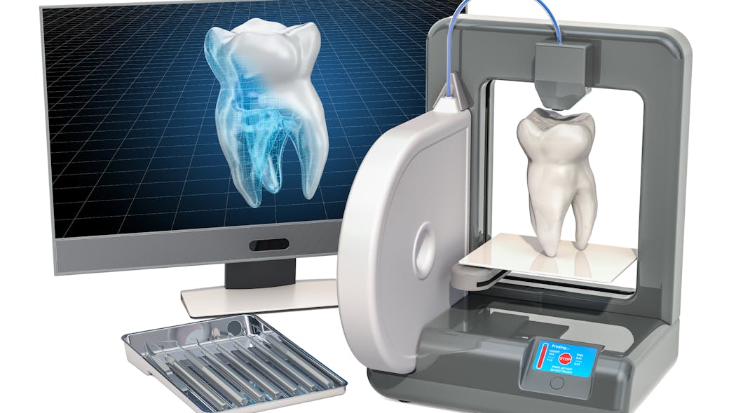
For almost 40 years,dentistry has been associated with some form of CAD/CAM technology. We have been scanning teeth and models for decades and using CNC technology to mill and manufacture restorations. This process has been purely reductive, meaning a block or ingot is ground or milled down to produce the final restoration. However, with the advent of additive manufacturing—think 3D printing—we are entering a new era of dental manufacturing, and it’s going to be a very exciting time.
Traditional milling of dental restorations began with diamond grinding of glass ceramics. Feldspathic blocks were ground using dual-motor CNC machines to create inlays/onlays and crowns. This process would sometimes take upward of 30–45 minutes. At the time, this was an incredible innovation, but today that amount of time would be simply unacceptable.
Fast-forward to today and the milling processes are light years ahead of where we started. Today, I can send a restoration from my CEREC Primescan, which was designed using only a few clicks, to my Primemill and have a full-contour zirconia restoration manufactured in less than five minutes. It’s incredible to think how much further we can go with the speed, accuracy, and efficiency we have in our current technology. And that is only in the chairside world.
Our laboratory partners have even more incredible technology that allows them not only to manufacture high-quality restorations efficiently, but on a much larger scale. Using large five-axis milling machines, laboratories can manufacture dozens of restorations at once out of a single puck of material. This has helped reduce the overhead for labs and increase their output. The modern dental lab technician may no longer have a CDT degree, but instead a computer science and graphic design background. Design and milling restorations will forever be a computer-guided process.
Within the last five to seven years, the world of 3D printing has exploded onto the dental scene. Formlabs was one of the first manufacturers to target the dental market with its Form2 printer. Using a vat filled with uncured resin, a very detailed laser would systematically cure the resin onto a build platform to create dental models from digital impressions. My first 3D printer would typically take 10–12 hours to manufacture a model, and that was completely acceptable at the time. In fact, it was exciting to finally eliminate alginate, stone, and a model trimmer!
Today, companies such as SprintRay and Dentsply Sirona are creating powerful and innovative 3D printing solutions that are speeding up the manufacturing process by leaps and bounds. On my Primeprint, I can produce a set of models in fewer than 30 minutes, which was unheard of just a few years ago.
With the rapid pace of innovation, research, and development—specifically regarding biocompatible materials and resins—it’s only a matter of time before additive manufacturing becomes the standard in dentistry. It took us almost 40 years to take a milled crown down from 40 minutes to five, and only two to three years to take a 12-hour printing process down to 30 minutes. We are already printing surgical guides and splints and provisionals today. I imagine within a few years, 3D printing of definitive restorations will have a significant place in the market. Regardless, I’m excited to just be along for the ride.
Editor’s note: This article appeared in the May 2022 print edition of Dental Economics magazine. Dentists in North America are eligible for a complimentary print subscription.
Which crown types are best for what situations?
View Image Gallery
Q: There is enormous confusion in the dental marketplace regarding which indirect restorations should be used and where in the mouth they should be used. The ads are not only confusing, but, in my opinion, they are also too optimistic. The significant commercial information claiming unprecedented success for specific restorations seems unrealistic. What is the state of the art for indirect restorations? What should be used and where in the mouth? How does heavy occlusion factor into the decision? Can optimum esthetics be achieved for all new materials? I would appreciate knowing what Clinical Research (CR) Foundation has found in long-term clinical studies.
A: I answered some of the same questions in “Which crown goes where?” but since then, many changes have occurred, more research is available, and questions keep coming up frequently. Importantly, the research on the various indirect restorations is starting to mature and provide some answers. In this article, I will give you a status report on the most-used crown types and their current success in CR/TRAC (Technologies in Restoratives and Caries Research Division of the CR Foundation) in vivo research. The information is divided into several locations in the mouth along with my suggestions as to the different strength and esthetic needs for those locations.
Also by Dr. Christensen:
Are endodontic posts really necessary?
The following overall statements relate to my answers.
- It is reported by large US dental labs that over 74% of indirect restorations are for single teeth.1
- Large labs report that ceramic indirect restorations currently comprise over 90% of the total indirect restorations made. The majority of those indirect restorations are milled from one of the zirconia variations, some are milled or pressed lithium disilicate, and a small number are conventional porcelain-fused-to-metal (PFM), or polymer.1
- A very conservative estimate is that about one-third of the adult population have either grinding or clenching bruxism, a highly important characteristic for restoration selection.
It can be concluded from the previous statements that different types of indirect restorations are present and that their various physical and esthetic characteristics relate to where they should be used. It is also important to note that additional long-term research is needed to confirm some of my suggestions.
Molars
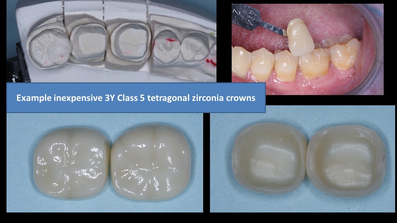
Optimum strength is available by using class 5, 3Y, tetragonal zirconia (figure 1). This is the original BruxZir formulation, now available from many Glidewell laboratories. Similar products are produced by other companies under other names. It is often called LT (“low translucency”) zirconia by dentists and labs. The strength and durability of this zirconia category have been confirmed through 11 years of in vivo research by the TRAC Division.

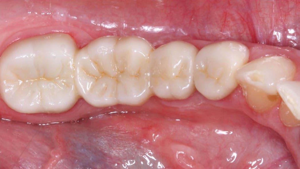
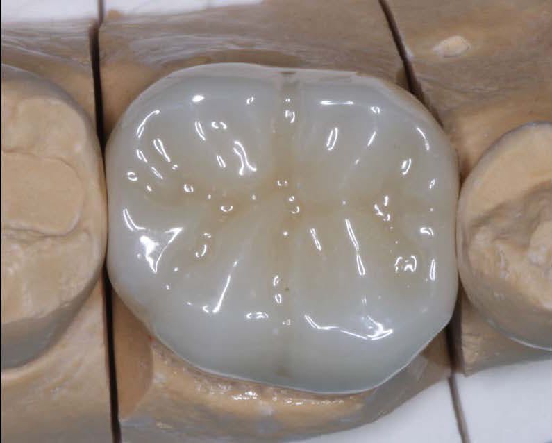
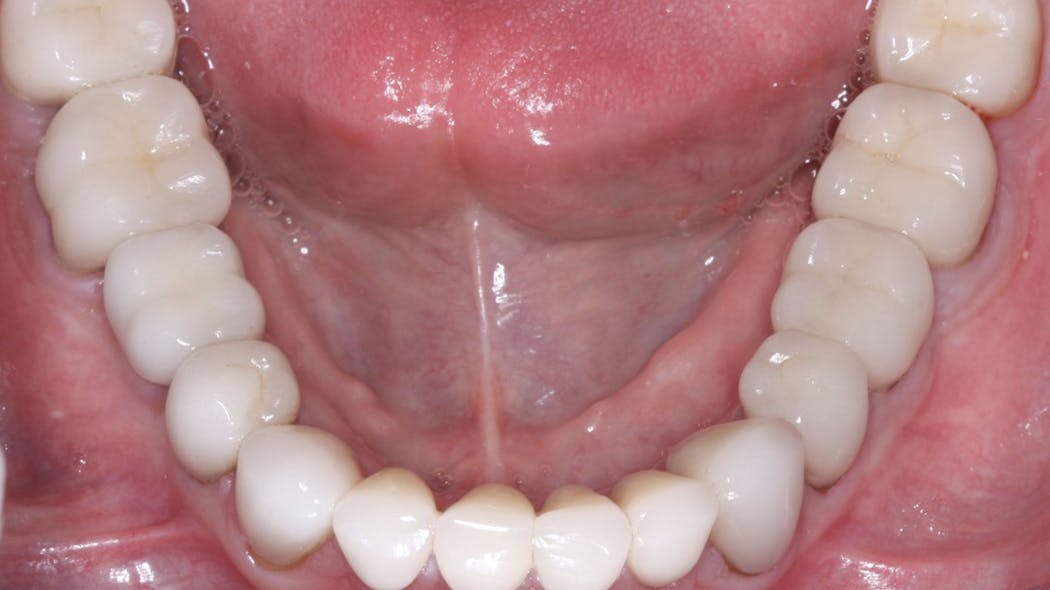
But, as most dentists know, this zirconia category has less-than-desirable esthetic qualities unless coated with layering ceramic or stained in the presintered zirconia stage (figures 2 and 3). Some dentists do not object to the unmodified color of this zirconia category for molars since it is not usually visible in the posterior of the mouth.
This zirconia formulation is well proven and has had unprecedented clinical success and lack of breakage. However, labs and manufacturers primarily promote the more esthetic forms of zirconia, identified as class 4 cubic zirconia. It is often described as HT (high translucency), or esthetic zirconia. This form of zirconia has lower strength than class 5 zirconia, and it still lacks long-term research for use in high-strength needs.
Currently, clinical research has mixed results, indicating promise for class 4 zirconia formulations but also some potential challenges. Since this formulation has been available for only a few years, you and your peers are doing much of the observational clinical research on these materials in your practices. Long-term clinical research is still needed to validate the use of this zirconia form for molars, bruxing patients, long-span fixed prostheses, and other high-strength clinical needs.
Premolars
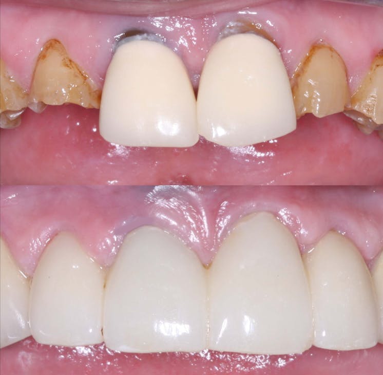
IPS e.max (lithium disilicate) is classified as a class 3 ceramic restoration. It is very well proven for single premolar restorations by both controlled studies and millions of such restorations placed internationally. As you know, it has unprecedented high esthetic qualities and strength (figures 4 and 5).

For nonbruxers, IPS e.max can be used safely for single premolars and select three-unit fixed prostheses involving both premolars and anterior teeth. It is suggested that at least 1.0 mm of IPS e.max thickness is present on all axial walls and 1.5–2.0 mm thickness on the occlusal surface for optimal strength. However, more fractures have been reported on multiple-unit lithium disilicate fixed prostheses in the premolar to anterior area than on single teeth.
Should IPS e.max be used on premolars in bruxing patients? Some practitioners are using it in bruxing situations because of its great success in nonbruxers. The only alternatives are porcelain-fused-to-metal (PFM) or class 4 zirconia. Selecting an appropriate restoration for a bruxing patient requires that the dentist have personal knowledge of the patient, the anticipated occlusal loading, and
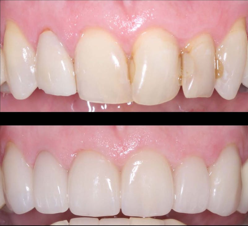
any esthetic needs.
Should class 4 cubic zirconia restorations be used for premolars? These modified zirconia forms are being highly promoted for such situations. Clinical observation by CR evaluators has been promising, but those observations are only short term. Developing challenges have already been noted in some brands by CR’s in vitro microscopic research, indicating the use of class 4 zirconia with caution until additional long-term research is available.
At present, IPS e.max is a well-proven product for single premolars and select three-unit fixed prostheses involving premolars.
In situations involving high-strength needs, such as in bruxing patients, color-modified class 5, 3Y zirconia is still a more-proven concept. Additional clinical research will determine if class 4 zirconia will be adequate for patients who are bruxers.
Anterior teeth
Anterior teeth usually have the least need for strength. The statements on premolars apply directly to anterior teeth, but esthetic acceptability is more important in the anterior area of the mouth.
If the restoration is for single teeth and the patient is not a bruxer, IPS e.max currently is the optimum restoration.
If a three-unit (or larger) fixed prosthesis is needed, color-modified class 5, tetragonal, 3Y zirconia may be optimum, especially for a bruxing patient, but such zirconia requires an artist/technician to achieve the best esthetics.
Should class 4 cubic zirconia be considered for anterior three-unit (or larger) fixed prostheses? The same challenges are present as those for premolars. More long-term research is needed. How long? At least several years of research on many brands. That research is beginning to come forth, but there are still numerous questions. If using class 4 zirconia for anterior restorations, I suggest observing the restorations carefully with high-power loupes at each recall appointment. Look for pits, minor cracks, excessive wear on opposing teeth, or other maladies. These challenges have been seen on some class 4 zirconia brands in preliminary research. It is our hope that class 4 zirconia brands will soon prove themselves in clinical research. In the meantime, be observant and cautious.
Summary
Significant confusion is present about what type of indirect restoration is best for specific situations. Current evidence, both scientific and observational, support the use of class 5, tetragonal, 3Y zirconia. However, this formulation has esthetic challenges that must be overcome. Class 4 cubic-containing zirconia has many formulations. Many brands are currently proving themselves, but more years will be necessary for that proof to be solidified.
IPS e.max is well proven for near universal use in nonbruxers and limited use in bruxers. In the meantime, don’t forget the more than 120 years of success dentistry has had with cast-gold alloy and the more than 65 years of success with porcelain-fused-to-metal.
The immediate future appears to point to continued and expanded use of zirconia indirect restorations with a slow reduction in the use of the excellent, well-proven IPS e.max.
Reference
- Based on data from Glidewell Labs.
Author’s note: The following educational materials from Practical Clinical Courses offer further resources on this topic for you and your staff.
One-hour videos:
- Cementing Restorations—Proven and Successful (Item no. 1921)
- Impressions Can Be Simple and Predictable (Item no. 1922)
Two-day hands-on courses in Utah:
- Restorative Dentistry 1—Restorative/Esthetic/Preventive with Dr. Gordon Christensen
- Faster, Easier, Higher Quality Dentistry with Dr. Gordon Christensen
For more information, visit pccdental.com or contact Practical Clinical Courses at (800) 223-6569.
Editor’s note: This article originally appeared in the February 2022 print edition of Dental Economics.
Fracture Resistance and Fracture Behaviour of Monolithic Multi-Layered Translucent Zirconia Fixed Dental Prostheses with Different Placing Strategies of Connector: An in vitro Study
Purpose: To evaluate the effect of different placing strategies performed in the connector area on fracture resistance and fracture behaviour of monolithic multi-layered translucent zirconia fixed dental prostheses (FDPs).
Materials and Methods: Thirty 3-unit monolithic FDPs were produced and divided into three groups (n = 10) based on the different strategies for placing the connector area of FDPs in multi-layered zirconia blank with varying contents of yttria ranging from 4 to 5 mol%. The groups were as follows: FDPs with connectors placed in dentin layer with 4 mol% yttria content, FDPs with connectors placed in gradient layer, and FDPs with connectors placed in translucent layer with 5 mol% yttria content. A final group (n = 10) of conventional monolithic zirconia with a monolayer of yttria content (4 mol%) has been used as a control group. The specimens were artificially aged using thermocycling and pre-loading procedures and subsequently loaded to fracture using a universal testing machine. Fracture loads and fracture behaviour were analyzed using one-way ANOVA and Fisher’s exact tests and statistically evaluated (p ≤ 0.05).
Results: There were no significant differences in fracture loads among the groups based on the placing strategies of the connector area of the FDPs in the multi-layered translucent zirconia blank (p > 0.05). There was no significant difference in fracture loads between monolithic multi-layered translucent zirconia and conventional monolithic translucent zirconia materials (p > 0.05). Fracture behaviour of FDPs with connector area placed in translucent layer differed significantly compared to FDPs with connector area placed in dentin layer and FDPs in control group (p = 0.004).
Conclusion: The placing strategies of the connector used in the computer aided design and manufacturing procedures do not considerably affect fracture resistance of monolithic FDPs made of multi-layered translucent zirconia. Monolithic FDPs made of multi-layered translucent zirconia show comparable strength to FDPs made of conventional translucent zirconia, but with different fracture behaviour.
Keywords: all-ceramic restorations, computer-aided design\manufacturing, fracture load, multi-layered zirconia, Y-TZP
Introduction
Yttria-stabilized tetragonal zirconia polycrystal (Y-TZP) is the most commonly used oxide ceramic material in Restorative Dentistry. This is related to its superior fracture strength and unique toughening properties.1,2 However, owing to its poor optical properties, Y-TZP based restorations must be veneered with translucent glass-ceramic materials in many clinical situations. Although the high success rate of veneered Y-TZP restorations has been reported to be over 90%, clinical complications such as veneer chipping and connector fracture still occur.3–5 Moreover, the use of veneered Y-TZP restorations requires removing more underlying tooth substance to provide enough space for the material. For those reasons, there is a general preference for shifting toward monolithic Y-TZP restorations, with challenges in achieving esthetical requirements without compromising the overall strength.6–8
The main drawback of using Y-TZP material as a monolithic restoration is the low translucency, resulting in poor esthetics.6,9–11 Scattering of light in Y-TZP and subsequent reduction of light transmittance mainly occurs at grain boundaries, pores, and secondary phases.6,9–11 However, enhanced optical properties of this material have been achieved by modifying the microstructure, for example, through altering the yttria (Y2O3) content and applying different sintering conditions.12,13 Shorter sintering times result in smaller grain size and thus an increase of the light transmittance of the final dental zirconia.12 Furthermore, it has been shown that the change of dopant contents, such as lanthanum oxide and aluminum oxide, improved the optical properties of zirconia.14 From a material point of view, the mechanical properties of Y-TZP are negatively affected by enhancing the translucent properties of the material.15,16 The more translucent the zirconia is, the lower the fracture strength.15,16
Recently, a new multi-layered translucent zirconia material, with a natural progression of shade and translucency, has emerged in the dental market to mimic natural teeth closely. This material is indicated to produce monolithic restorations in both the anterior and posterior regions. There are two types of multi-layered translucent zirconia on the market: 1) Multi-layered zirconia with different colour saturations in the different layers but the same yttria content throughout all layers, and 2) Multi-layered zirconia with different translucency in the different layers as a result of varying yttria contents in the different layers. Thus, the strength and toughness of the layers with different yttria contents are expected to be different. During computer-aided design and manufacturing (CAD/CAM) procedures, dental technicians can use different placing strategies to place the fixed dental prosthesis (FDP) in multi-layered translucent zirconia blank before milling. Previous studies showed that the main fracture origin leading to the failure of the prostheses is located at the gingival side of the connector area, which is linked to the development of stress concentrations in the connector when different loads are applied to the FDPs.17,18 Accordingly, in practice, the fracture resistance of the FDP, especially in the connector area, might be affected depending on how the placing strategy has been performed by the dental technician during CAD/CAM procedures. It is not known, however, if the different placing strategies of the connector, during computer manufacturing of zirconia blanks, might affect the fracture resistance of the final restoration made of the new multi-layered translucent zirconia material, since the strength varies between the different layers of zirconia.
Therefore, the present study aimed to evaluate the effect of the different placing strategies performed in the connector area on fracture resistance and fracture behaviour of monolithic FDPs made of multi-layered translucent zirconia. The null hypothesis is that there is no difference in fracture resistance and fracture behaviour of the FDPs made of multi-layered translucent zirconia based on the placing strategies performed in the connector area.
Materials and Methods
Study Design
Thirty 3-unit monolithic FDPs were produced and divided into three groups (n=10) according to the different strategies for placing the connector area of the FDPs in the multi-layered translucent zirconia blank (IPS e.max ZirCAD MT Multi, Ivoclar Vivadent, Schaan, Liechtenstein) (Figure 1). The groups were as follows: FDPs produced with the connectors placed in the dentin layer with 4 mol% yttria, FDPs produced with the connectors placed in the gradient layer, and FDPs produced with the connectors placed in the translucent layer with 5 mol% yttria. A final group (n=10) of conventional monolithic zirconia with monolayer of 4 mol% yttria content has been used as a control group (IPS e.max ZirCAD, Ivoclar Vivadent, Schaan, Liechtenstein). The FDPs were cemented using compatible resin cement onto abutment models made of a polymer material (POM C glass infiltrated). The specimens were artificially aged using both thermocycling and cyclic fatigue procedures before they were loaded to fracture. Fracture loads and fracture behaviour were subsequently analyzed and evaluated statistically p ≤0.05.
Specimen Preparation
For the preparation of the teeth, a plastic model of a mandibular jaw was used (KaVo YZ; KaVo Dental GmbH, Biberach, Germany). The preparations were made on the canine (43) and premolar (45) and were designed to provide space for Y-TZP material with a 120° chamfer and 15° convergence angle. The teeth preparations were conducted by prosthodontist. After the preparations were conducted, a full-arch impression using silicone material (President; Coltene AG, Altstätten, Switzerland) was made and poured with die stone material (Vel-Mix; Kerr Corp, Orange, CA). A master cast was produced from the die stone, and subsequently, a wax-up (1.5–3 mm) of the FDP was made by professional dental technician. The wax-up was scanned with a double-scan technique using a dental laboratory scanner (D900L; 3Shape, Copenhagen, Denmark). Data from the scanner were transferred to a computer loaded with computer-aided design (CAD) software. The design of the FDP connector was a round shape and the dimensions for all the FDP connectors were adjusted to 3 mm x 3 mm. The occlusal thickness of the retainer core was set to 1 mm, and the axial wall thickness was set to 0.8 mm with a 0.5 mm cervical margin. After the adjustments, the CAD file was sent to a milling center (Cosmodent AB, Malmö, Sweden) to produce the FDPs. The same sintering protocol for the two zirconia materials has been used following the manufacturer instructions. The CAD file was used to produce the abutment models made from a polymer material (POM-C GF25; Mekaniska AB, Simrishamn, Sweden) with a modulus of elasticity comparably close to dentin (9 GPa).
Artificial Aging, Cementation, and Load to Fracture Test
All FDPs were subjected to artificial aging, beginning with thermocycling. In a thermocycling device (THE-1100; SD Mechatronik GmbH, Feldkirchen-Westerham, Germany) containing two water baths, the FDPs underwent 10,000 thermocycles at two different temperatures, 5 and 55°C. Each cycle lasted for 60 seconds, 20 seconds in each bath and 20 seconds to complete the transfer between the baths.4,19–22 The cementation of the FDPs to the abutment models was completed using a dual-polymerized resin cement (Panavia V5; Kuraray Medical Inc., Okayama, Japan) according to the manufacturer’s recommendations. However, before cementation, the abutment models were air-abraded with 50 µm aluminum oxide using an air abrasion device (Basic Quattro IS; Renfert GmbH, Hilzingen, Germany) as well as treated with two primers (Tooth Primer, Clearfil and Ceramic Primer; Kuraray Medical Inc) following the manufacturers’ instructions. The FDPs were cemented to the abutment models with a standardized seating load of 15 N in the direction of insertion. A calibrated curing lamp (Heraeus Translux® Power Blue®, Heraeus Kulzer GmbH) was used according to the manufacturer’s recommendations to initiate the curing. Ultimately, excess cement was removed with a scalpel (AESCULAP® no. 12, Aesculap AG & Co, Tuttlingen, Germany). The specimens were stored in a humid environment at a temperature of 37°C before cyclic fatigue. The last step of artificial aging was cyclic fatigue using a pre-loading machine (MTI Engineering AB; Lund, Sweden/Pamaco AB, Malmö, Sweden). The cemented FDPs were submerged in distilled water at 10° of inclination towards the tooth axis and went through 10,000 cycles of 30–300 N at a frequency of 1 Hz. A 4 mm stainless ball was placed on the occlusal surface of the connector area between teeth 45 and 44 of the bridges to apply mechanical cyclic loads.4,19–22
After artificial aging, all FDPs were installed in a test jig at 10° inclination towards the axial direction using a universal testing machine (Instron 4465, Instron Co. Ltd, Norwood, MA, USA), (Figure 2) as was suggested in previous laboratory studies.4,19–22 The load was applied on the pontic using a specialized stainless-steel intender. Throughout loading, all the FDPs were submerged in water. The crosshead speed was set at 0.255 mm/min, and the fracture was defined as follows: visible crack, load drop or an acoustic event, whatever occurred first.4,19–22 The load at fracture was then registered.
| Figure 2 Illustration of the specimen in a test jig at 10° inclination in cyclic fatigue and load to fracture tests. All specimens were submerged in water during the tests. |
Fracture Behaviour Analysis
The fracture surfaces of the FDPs were analyzed by two examiners. A gross visual and microscopic assessments (Leica DFC 420, Leica Application Suite v. 3.3.1, Leica Microsystems CMS GmbH, Wetzlar, Germany) were performed to classify fracture behaviour according to the location of fracture into: fracture at the distal connector, fracture at the mesial connector, complete fracture of the FDP (involving fracture of the retainer).
Statistical Analysis
The differences in fracture resistance among the groups were analyzed using one-way ANOVA, followed by Tukey’s post hoc test (IBM SPSS Statistics 25). The differences in fracture behaviour among the groups were analyzed using Fisher’s exact test. The level of significance was set to p ≤0.05. The statistical analysis was performed by an experienced professional statistician. Power analysis was based on previous studies where differences regarding the level of significance and standard deviation were detected among the zirconia-based specimens.17,19–21
Results
Loads at fracture, levels of significance, fracture behaviour for all groups are summarized in Tables 1 and 2. There were no significant differences in fracture loads among the groups based on the different strategies for placing the connector area of the FDPs in the multi-layered zirconia blank (p >0.05). There was no significant difference (p >0.05) in fracture loads between the two different materials: monolithic multi-layered translucent zirconia and conventional monolithic translucent zirconia materials.
Three types of fracture behaviour were registered after load to fracture test: fracture at the mesial connector propagating through the pontic, fracture at the distal connector propagating through the pontic, and complete fracture involving the retainer (Figure 3). Fracture behaviour of the FDPs with connector area placed in the translucent layer (5Y-TZP) differed significantly compared to the FDPs with connector area placed in the dentin layer (4Y-TZP) and the FDPs in the control group (p ≤0.05).
| Figure 3 Different types of fracture behaviour. (A) Fracture at distal connector; (B) complete fracture; (C) fracture at mesial connector. |
Discussion
The null hypothesis of this study was rejected since fracture resistance of the FDPs showed no significant differences among the groups based on the different placing strategies performed in the connector area during computer manufacturing of the FDPs. However, the results showed that the different placing strategies performed in the connector area affect fracture behaviour of the three-unit FDPs.
One of the common methods to improve the translucency of dental zirconia is by changing the amount of yttria content, which results in a greater portion of the optically isotropic cubic phase without light scattering at the grain boundaries.14,15The major phenomena related with the enhanced translucency of polycrystalline zirconia-based ceramics is the reduction of birefringence, the light scattering promoted by a material with anisotropic refractive index. Tetragonal zirconia phase is birefringent, however, by increasing yttria content the precipitation of cubic zirconia, which is isotropic and do not experience birefringence, is favoured and an enhancement of the transmitted light fraction is experienced.23–25 This, on the other hand, compromises the strength and toughness of the cubic zirconia because it does not undergo stress-induced transformation.14,23–25 In the present study, the FDPs made of multi-layered translucent zirconia were divided into three groups: dentin, gradient, and translucent, based on the content of yttria ranging from 4 to 5 mol%. The groups with the connectors placed in the gradient and the translucent layers presented higher standard deviation values than the dentin and control groups. This might be explained by the fact that the gradient layer combines different microstructures of both the translucent and the dentin layers, which results in varying mechanical properties. Thus, the FDPs with the connectors placed in the layer consisting of a microstructure primarily composed of dentin (4Y-TZP) withstand higher fracture loads. The opposite applies to the FDPs with the connectors placed in the layer consisting of a mainly translucent microstructure, namely 5Y-TZP. These findings are in line with previous studies, which concluded that translucency affects the mechanical properties of zirconia.15,23–25 Although the differences of the results were not statistically significant, the numerical differences among the groups in this study, together with the findings of previous studies, confirm the effect of enhanced translucency on the mechanical properties of Y-TZP. Moreover, it is noteworthy that the limitations of the methodology used in this study might have influenced the results. For geometric reasons, it is impossible to place the whole reconstruction in one layer in the multi-layered translucent zirconia blank without infringing the minimum dimensional demands of the FDP. This means that the critical part of the connector area, the gingival portion, where the highest stress concentrations occur during loading, will probably not be entirely located in solely one layer.17,18 This technical limitation means that study findings need to be interpreted cautiously.
Many studies have investigated the adverse effects of the other methods of enhancing the optical properties of zirconia on mechanical properties. For instance, although doping of metal oxides improves the optical properties of zirconia, this may affect adversely the mechanical and biological (cytotoxic) properties of zirconia.14 Other fabrication techniques such as colouring of pre-sintered zirconia for enhancing the optical properties might be necessary in many clinical cases. Previous studies have shown the effect of such colouring techniques on the mechanical and optical properties.26,27 Nevertheless, a very recent study investigating new multi-layered translucent zirconia material showed no differences in neither microstructure nor translucency between the different layers.28 Only colour pigment composition is different between the layers within each multi-layered translucent zirconia blank. The same study revealed that lanthanum oxide doping improved the translucency without diminishing the mechanical properties of the multi-layered translucent zirconia, which is the main goal when developing high esthetical monolithic dental zirconia.
Considering fracture behaviour, this study showed that most fractures started from the connector area (mesial or distal) and propagated through the pontic during loading. This is in agreement with previous studies, which concluded that critical tensile stresses mostly develop in the gingival embrasure of the connector, result in failure of prosthesis.17,18 However, there were significantly more complete fractures (involving the retainer) in the FDPs with connector area placed in the translucent layer (5Y-TZP) compared to the FDPs of the other groups. This finding could be expected theoretically since the translucent layer has a microstructure that is less resistant to fractures, as previous studies have shown.15,23–25 It is noteworthy that fracture behaviour analysis in this study aimed to show the fracture initiation and propagation pattern under a light microscope and evaluate the ability of the test to mimic the clinical failures of dental restorations. Sophisticated fractographic analysis using a scanning electron microscope, however, might provide more details on fracture behaviour.
When conducting an in vitro study to evaluate the mechanical properties of new material, a laboratory setup simulating the oral environment and the complex forces of mastication is of great importance. One of the limitations of in vitro studies is the difficulty to choose which aging procedures would produce comparable clinical results. Previous studies have investigated the effect of artificial aging procedures, that used to mimic the clinical situation, on the longevity of ceramics. Despite that some of those studies fail to show a direct relationship between aging procedures and fracture resistance of ceramics,29 most agree that they have a significant effect on the longevity of ceramic materials.30–32 Therefore, there is no consensus regarding the effectiveness of aging tests or a specific aging protocol, but it was reasonable, however, to perform such procedures in the present study to allow for comparison of the results of other studies carried out by the same research group with this specific protocol.4,19–21 The FDPs were mounted with a 10 of inclination relative to the load direction in the load to fracture test. This angle of inclination has been used in many previous studies and was initially suggested by Yoshinari and Derand.4,19–22 However, the mechanical load to fracture test performed in a laboratory study can never completely reproduce loads and environmental influences as in the clinical situation but can still give important information. Furthermore, to obtain realistic fracture load values and compare these values with previous studies, replicating the real clinical situation concerning mechanical support is crucial.33 Therefore, all FDPs were cemented onto abutment models made of a material with a modulus of elasticity close to dentin. The cementation procedure was performed according to the manufacturer’s recommendations, and the same cement was used for all groups. Since in vitro studies have shown that thermocycling affects the bond strength of cements, all FDPs were cemented after this stage to avoid partly loose prostheses at the subsequent cyclic fatigue and load to fracture tests.31,32
Since adequate communication between the dentist and the dental technician is essential for successful dental restorations, it is a prerequisite for dentists to gain knowledge of the dental material that is required. This study has shown that the different strategies for placing the FDP in the blank during the CAD/CAM process do not have a critical effect on the mechanical properties of the translucent multi-layered zirconia FDPs. Thus, this facilitates the process of ordering for the dentist who does not have to pay regard to the technical aspects. In vitro studies, in line with the present one, are of great importance to evaluate new dental materials before using them in a clinical situation, thus safeguarding patient safety.
Conclusion
Within the limitations of this laboratory study, the following conclusions can be drawn: the placing strategies of the connector used in the computer aided design and manufacturing procedures do not considerably affect fracture resistance of monolithic FDPs made of multi-layered translucent zirconia. Monolithic FDPs made of multi-layered translucent zirconia show comparable strength to FDPs made of conventional translucent zirconia, but with different fracture behaviour.
Improving the color match of zirconia crowns
QUESTION:
Most of the zirconia crowns and fixed prostheses coming from my labs lately are too light in color, despite my providing preoperative photos and descriptions to the technicians. I have tried several different laboratories, and I am still frustrated with the mismatched colors. Although zirconia crowns are strong, I believe their color-matching is severely lacking. My colleagues have expressed similar color mismatch as well. What can be done to improve this obvious problem?
ANSWER:
I agree completely with your criticism of the current generation of zirconia crowns. The Technologies in Restoratives and Caries Research (TRAC) Division of the Clinical Research Foundation has accomplished long-term in vivo research on zirconia crowns and found their strength to be fantastic and their service over many years to be outstanding. However, only recently has the color of zirconia crowns been improved so they are more acceptable.
The changes to improve color and translucence of the crowns has been successful, but it also has had some negative effects. The information in Figure 1 is from Glidewell Laboratories and shows remarkable changes over the last several years as ceramic restorations have become mainstream. What are manufacturers doing to improve the esthetics of zirconia restorations? To answer that question, let’s discuss the various types of zirconia.
Zirconia and its various forms
When observing the 2020 data from Glidewell Laboratories in Figure 1, it is obvious that zirconia wins the popularity contest. As you probably know, Glidewell started this clinical revolution with BruxZir, which soon dominated the market. Zirconia-based crowns have been available for more than 21 years, and full-zirconia crowns have been available for about 10 years.
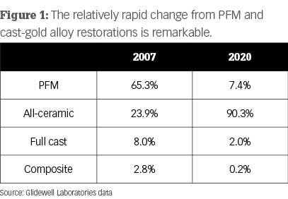
In the first few production years, the original zirconia-based crowns had challenges, but since then those problems have largely been overcome. Despite that, the use of zirconia-based restorations has not grown markedly.
Full-zirconia restorations of various formulations are extremely popular and state-of-the-art. But zirconia is not zirconia. You are undoubtedly familiar with the ADA/ISO classification of ceramic crowns, which we previously published in the July 2019 issue of Clinicians Report. (Visit cliniciansreport.orgto read the article in detail.)
The initial BruxZir formulation was classified as class 5 tetragonal zirconia. This is probably what you will receive if you order class 5 or 3Y (3 molar percent yttrium oxide) tetragonal zirconia. This formulation has been used in nondental applications in various industries for more than 30 years. It is the strongest form of zirconia and has optimal transformation toughening (lack of crack propagation) during service. Unfortunately, this is the iteration of dental zirconia most difficult to make esthetically acceptable.
If you are planning a long-span fixed partial denture (FPD) or treating a patient with bruxism, this is an excellent choice shown to be comparable in strength to porcelain-fused-to-metal (PFM) in our long-term TRAC studies. It can be made esthetically acceptable for some situations by presinter staining or optimum use of external stains.
Manufacturers have been experimenting with making 3Y zirconia more esthetic. The most successful and easiest method to date is to increase the translucence by adding more translucent material to the zirconia. ADA/ISO classifies this type of zirconia as class 4 cubic-containing zirconia. It is also identified as 4Y or 5Y or combinations of those percentages. Although additional long-term clinical research is still needed for the so-called esthetic zirconia, class 4 zirconia currently appears to be adequate for singles, short-span FPDs, and other situations not requiring the strength of 3Y zirconia.
Why do you need to know the zirconia classifications?
Your knowledge of these classifications allows you to communicate with salespeople and laboratory technicians, as well as understand what materials you are putting into your patients’ mouths. Knowing this, you’ll better understand the changes that are being made in the original dental zirconia restorations.
Adding more translucent materials to the zirconia. Additional oxides are now being added to the original Glidewell formulation of zirconia (3Y, class 5 zirconia versions) by many manufacturers. This formulation change improves translucency and esthetics. However, the change to 4Y or 5Y or combinations of those reduces the overall strength of zirconia and its ability to “heal” microcracks. (Higher transformation toughening increases the lack of subsequent crack propagation.) Although this procedure reduces strength, clinical success is currently being proven and reported in the field by practicing dentists. The esthetic characteristics of some of these class 4 restorations can rival IPS e.max (Ivoclar Vivadent).
Placing relatively thick layers of stain and glaze on the external of the restorations. Many labs are firing ceramic on the outside of the mismatched zirconia to improve the zirconia color (figure 2). In our research, these additions are usually feldspathic ceramic and are showing wear on the opposing teeth. There is also a slow but continuing loss of the added superficial layers. If the external layers are thin, the result is a slow change back to the original zirconia colors.
Internal staining of zirconia. Some labs are staining the 3Y, class 5 zirconia in the presintered stage, which improves the color significantly. However, this requires artistic technicians, more time, and a greater cost to the dentist (figure 3).
Currently, most indirect restorations are ceramic, and that trend will undoubtedly continue to grow. Metal and porcelain-to-metal restorations are showing a relatively rapid decrease in use in dentistry.
Is there a dental laboratory problem?
Yes! The laboratory industry has changed rapidly and significantly. Until recently, porcelain-fused-to-metal restorations were the norm. That situation then changed to nearly all ceramic restorations, with zirconia restorations being rapidly produced by milling machines that use computer-driven software and by lithium disilicate being pressed or milled.
Many artistic laboratory technicians are being replaced with highly competent, computer-savvy technicians who produce restorations at record speeds considered to be impossible by older technicians. As a result, restorations are less expensive but often less esthetically acceptable.
If needed, clinicians must seek artistically oriented laboratory technicians to accomplish the complex esthetic results desired for some patients, and consequently, we must expect to pay higher fees for these restorations. Esthetic restorations are available—certainly with lithium disilicate (IPS e.max) and, with effort, even zirconia.
Which indirect restorations are desirable and for what situations?
I have heard some dentists comment with mixed opinions on the esthetic dilemma about which you asked. Most dentists admit, to an embarrassing degree, that many zirconia restorations do not adequately match adjacent teeth, but they further comment that zirconia restorations in the posterior nonesthetic regions are far more esthetically acceptable than gold alloy or porcelain-fused-to-metal when the glaze and staining have worn off.
Porcelain-fused-to-metal
Don’t forget the 70-year success of these restorations, especially when patients have had proven success with previous PFM restorations in their mouths. The advantages and disadvantages of these restorations are well known. Many labs can make highly esthetic PFM restorations. They are especially useful in clinical situations requiring precision or semiprecision attachments, which are not currently available for zirconia or lithium disilicate restorations. An additional continuing use for PFM is long-span fixed prostheses. Although not as commonly used as in the past because of the availability of implants, PFM restorations are still occasionally needed.
Lithium disilicate (most common brand name IPS e.max)
Can you name any other proven successful type of indirect restoration that equals the superior esthetic result of e.max for single-tooth restorations? That question is easy to answer. Many crown types have been tried and were initially successful, but they failed the need for longevity. The success of e.max in selected three-unit anterior FPDs has been shown, but I suggest that the newer generations of zirconia (class 4 zirconia) will probably prove to be more successful for long-term clinical success in these anterior situations.
Summary
The crown revolution has changed almost every treatment plan that requires crowns or fixed prostheses. This article describes the state-of-the-art and makes suggestions about which materials to use, their advantages and limitations, as well as how to produce the esthetic result necessary for specific clinical situations.
Author’s note: The following educational materials from Practical Clinical Courses offer further resources on this topic for you and your staff.
